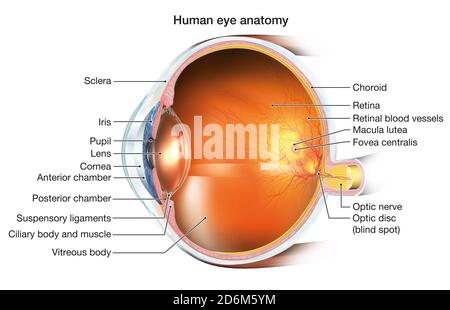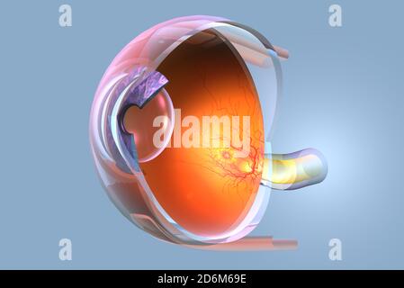
Medically 3D illustration showing human eye with retina, pupil, iris, anterior chamber, posterior chamber, ciliary body, eye ball, blood vessels, macu Stock Photo
RF2D6M69E–Medically 3D illustration showing human eye with retina, pupil, iris, anterior chamber, posterior chamber, ciliary body, eye ball, blood vessels, macu
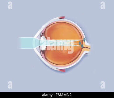
Anatomy of human eye showing focal points. Stock Photo
RME8MMN2–Anatomy of human eye showing focal points.
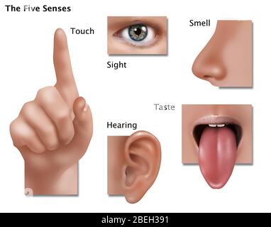
An illustration showing the five senses: touch, sight, smell, taste, and hearing. Stock Photo
RM2BEH391–An illustration showing the five senses: touch, sight, smell, taste, and hearing.
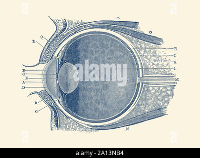
Vintage anatomy print showing a diagram of the human eye. Stock Photo
RF2A13NB4–Vintage anatomy print showing a diagram of the human eye.
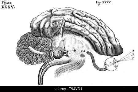
Historical diagram of the brain, showing the process of sight, by Rene Descartes. The Nervous System, 1662. Stock Photo
RMT94YD1–Historical diagram of the brain, showing the process of sight, by Rene Descartes. The Nervous System, 1662.
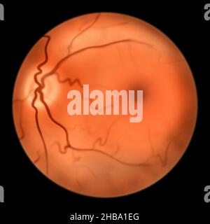
Illustration of the healthy eye retina showing optic disk, blood vessels, macula, and fovea. Stock Photo
RF2HBA1EG–Illustration of the healthy eye retina showing optic disk, blood vessels, macula, and fovea.
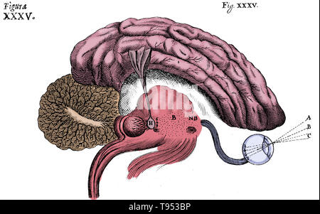
Historical diagram of the brain, showing the process of sight, by Rene Descartes. The Nervous System, 1662. Stock Photo
RMT953BP–Historical diagram of the brain, showing the process of sight, by Rene Descartes. The Nervous System, 1662.
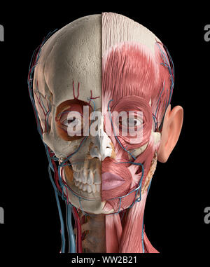
Human head anatomy 3d illustration. Showing skull, facial muscles, veins and arteries. On black background. Stock Photo
RFWW2B21–Human head anatomy 3d illustration. Showing skull, facial muscles, veins and arteries. On black background.
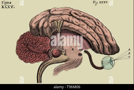
Historical diagram of the brain, showing the process of sight, by Rene Descartes. The Nervous System, 1662. Stock Photo
RMT96M06–Historical diagram of the brain, showing the process of sight, by Rene Descartes. The Nervous System, 1662.
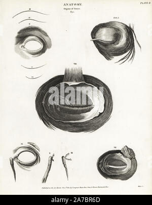
Anatomy of the human eye, showing musculature, tear duct, etc. Copperplate engraving by Milton from Abraham Rees' Cyclopedia or Universal Dictionary of Arts, Sciences and Literature, Longman, Hurst, Rees, Orme and Brown, London, 1820. Stock Photo
RM2A7BR6D–Anatomy of the human eye, showing musculature, tear duct, etc. Copperplate engraving by Milton from Abraham Rees' Cyclopedia or Universal Dictionary of Arts, Sciences and Literature, Longman, Hurst, Rees, Orme and Brown, London, 1820.
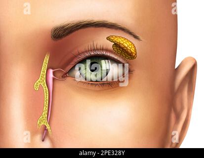
Anatomy drawing showing the functioning of lacrimal glands. Digital illustration. Stock Photo
RF2PE328Y–Anatomy drawing showing the functioning of lacrimal glands. Digital illustration.
Medically 3D illustration showing human eye with intraocular lens (IOL), retina, pupil, iris, anterior chamber, posterior chamber, ciliary body, eye b Stock Photo
RF2D6M5YM–Medically 3D illustration showing human eye with intraocular lens (IOL), retina, pupil, iris, anterior chamber, posterior chamber, ciliary body, eye b