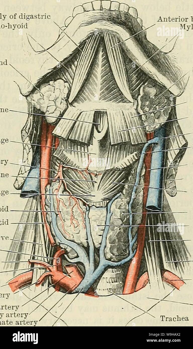
Archive image from page 1420 of Cunningham's Text-book of anatomy (1914). Cunningham's Text-book of anatomy cunninghamstextb00cunn Year: 1914 ( THE NECK. 1387 with the carotid sheath and its contents, being retracted well forwards. In approach- ing the trunk of the inferior thyreoid artery from the front the sterno-mastoid and carotid sheath are retracted backwards and the dissection is continued through the cellular interval between it and the sheath (outer capsule) of the thyreoid gland, which is formed by the splitting of the pretracheal fascia. A glandular abscess in this compartment usua
Image details
Contributor:
Bookive / Alamy Stock PhotoImage ID:
W9HAX2File size:
5.7 MB (359.9 KB Compressed download)Releases:
Model - no | Property - noDo I need a release?Dimensions:
1088 x 1839 px | 18.4 x 31.1 cm | 7.3 x 12.3 inches | 150dpiMore information:
This image is a public domain image, which means either that copyright has expired in the image or the copyright holder has waived their copyright. Alamy charges you a fee for access to the high resolution copy of the image.
This image could have imperfections as it’s either historical or reportage.
Archive image from page 1420 of Cunningham's Text-book of anatomy (1914). Cunningham's Text-book of anatomy cunninghamstextb00cunn Year: 1914 ( THE NECK. 1387 with the carotid sheath and its contents, being retracted well forwards. In approach- ing the trunk of the inferior thyreoid artery from the front the sterno-mastoid and carotid sheath are retracted backwards and the dissection is continued through the cellular interval between it and the sheath (outer capsule) of the thyreoid gland, which is formed by the splitting of the pretracheal fascia. A glandular abscess in this compartment usually points upon the surface, adhesions being formed, first, between the gland and the fascia, and, subsequently, between the latter and the cutaneous structures. In diffuse suppurative cellulitis of this compartment the pus burrows towards the root of the neck, and may reach either the mediastinum or the axilla. Medial Line of the Neck.—The body of the hyoid bone divides the median plane of the neck into supra- and infra-hyoid portions. Above the hyoid bone is the sub- mental triangle, with its apex at the inferior border of the symphysis menti and its Anterior belly of digastric Mylo-hyoid Submaxillary gland Fig. Tbyreo-hyoid membrane Thyreoid cartilage Superior thyreoid artery Crico-thyreoid membrane Cricoid cartilage Lateral lobe of thyreoid Common carotid —j Phrenic nerveT~J-_j Inferior thyreoid Transversalis colli Vertebral artery Subclavian artery Transverse scapular artery Common carotid artery ' / Internal mammary artery' /~V Innominate artery / Inferior thyreoid vein 1087.—Dissection of the Front of the Neck. have been removed, and the lower part of the right parts (from Cunningham). Anterior belly of digastric Mylo-hyoid Submaxillary gland Omo-hyoid Sterno-hyoid Internal jugular vein Superior thyreoid vein Common carotid artery Sterno-mastoid Crico-thyreoid muscle Lateral lobe of thyreoid body Isthmus of thyreoid Scalenus anterior Scalenus medius Subclavia