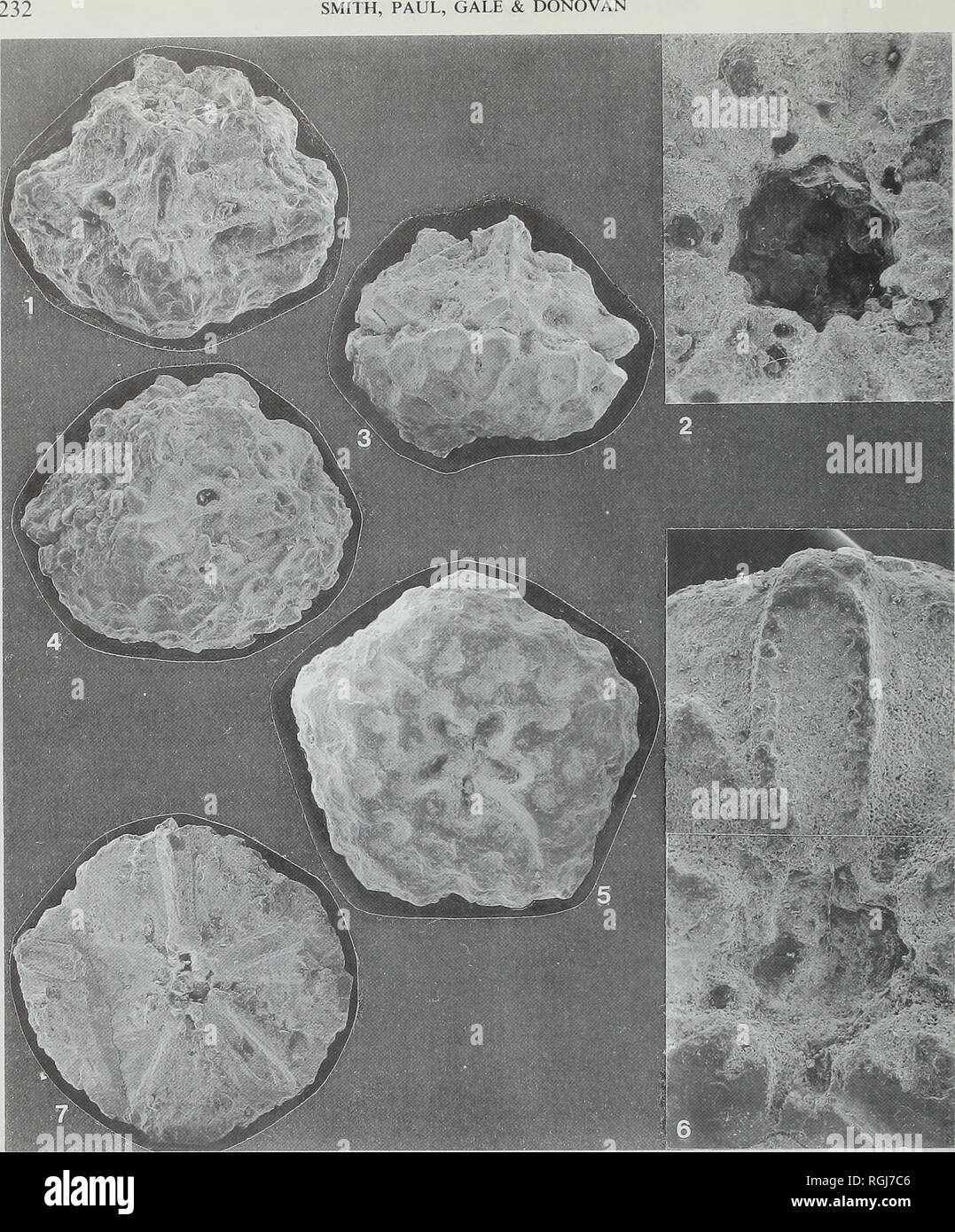
. Bulletin of the British Museum (Natural History), Geology. 232. Plate 50 Glenotremites cf. aequimarginatus (Carpenter) Fig. 1 E.69474, oblique lateral view centred on an interradius (x 91). Note the deep groove along the radial: radial suture. See also PI. 51, figs 1-3. Fig. 2 E.69473, detail of the central cavity on the centrodorsal (x 22). Note the radial nerve canals around the central cavity. Fig. 3 E.69479, oblique view of centrodorsal showing the cirral facets ( x 8). See also PI. 51, figs 4-5. Fig. 5 E.69477, dorsal view of a centrodorsal showing a large dorsal star ( x 8). See also P
Image details
Contributor:
Book Worm / Alamy Stock PhotoImage ID:
RGJ7C6File size:
7.1 MB (573.2 KB Compressed download)Releases:
Model - no | Property - noDo I need a release?Dimensions:
1439 x 1736 px | 24.4 x 29.4 cm | 9.6 x 11.6 inches | 150dpiMore information:
This image is a public domain image, which means either that copyright has expired in the image or the copyright holder has waived their copyright. Alamy charges you a fee for access to the high resolution copy of the image.
This image could have imperfections as it’s either historical or reportage.
. Bulletin of the British Museum (Natural History), Geology. 232. Plate 50 Glenotremites cf. aequimarginatus (Carpenter) Fig. 1 E.69474, oblique lateral view centred on an interradius (x 91). Note the deep groove along the radial: radial suture. See also PI. 51, figs 1-3. Fig. 2 E.69473, detail of the central cavity on the centrodorsal (x 22). Note the radial nerve canals around the central cavity. Fig. 3 E.69479, oblique view of centrodorsal showing the cirral facets ( x 8). See also PI. 51, figs 4-5. Fig. 5 E.69477, dorsal view of a centrodorsal showing a large dorsal star ( x 8). See also PI. 51, fig. 8. Glenotremites rotundus (Carpenter) Fig. 4 E.69481, lateral view showing hemispherical profile of centrodorsal and well preserved bra- chial facet (x 8-7).. Please note that these images are extracted from scanned page images that may have been digitally enhanced for readability - coloration and appearance of these illustrations may not perfectly resemble the original work.. British Museum (Natural History). London : BM(NH)