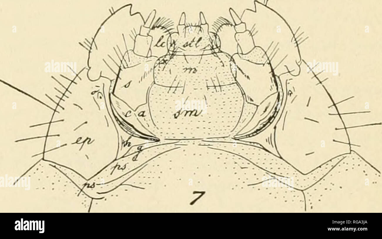
. Bulletin of the U.S. Department of Agriculture. Agriculture. Egg and Larva of the Tobacco Beetle (Lasioderma serricorne). Fig. 1. -Front view of face. Fig. 2.—Side view of larva. Fig. 3.—Thoracic and abdominal areas: aa, Parasternum; a, labial palp; ae,alar area; b,stipes labialis; c,mentum; d,submentum; <ll, dorso- lateral suture; e, gula:/, presternite; g,antenna; h, ocellus; i, stipesmaxillaris; k, cardo; /, epistoma; m, clypeus; m,labrum: o.preepipleurum; oe, basisternum; p, epipleural lobe; g, postepipleurum; r, prehypopleurum: s, posthypopleurum; r.prescutum; «, scutum; v, scutellum
Image details
Contributor:
Book Worm / Alamy Stock PhotoImage ID:
RGA3JAFile size:
7.1 MB (216.3 KB Compressed download)Releases:
Model - no | Property - noDo I need a release?Dimensions:
2118 x 1179 px | 35.9 x 20 cm | 14.1 x 7.9 inches | 150dpiMore information:
This image is a public domain image, which means either that copyright has expired in the image or the copyright holder has waived their copyright. Alamy charges you a fee for access to the high resolution copy of the image.
This image could have imperfections as it’s either historical or reportage.
. Bulletin of the U.S. Department of Agriculture. Agriculture. Egg and Larva of the Tobacco Beetle (Lasioderma serricorne). Fig. 1. -Front view of face. Fig. 2.—Side view of larva. Fig. 3.—Thoracic and abdominal areas: aa, Parasternum; a, labial palp; ae, alar area; b, stipes labialis; c, mentum; d, submentum; <ll, dorso- lateral suture; e, gula:/, presternite; g, antenna; h, ocellus; i, stipesmaxillaris; k, cardo; /, epistoma; m, clypeus; m, labrum: o.preepipleurum; oe, basisternum; p, epipleural lobe; g, postepipleurum; r, prehypopleurum: s, posthypopleurum; r.prescutum; «, scutum; v, scutellum; W, ventrolateral suture; x, postscutellum. Fig. 4.—Anterior portions of face (labrum, clypeus, epistoma, and frons): Ant, antenna; c, clypeus; cl, epistoma, lateral part; em, epistoma, median part;/, frons. Fig. 5.— Prothoracic leg. Fig. 6.—Mandible, ventral side. Fig. 7.—Underside of head: a, Maxillary articu- lating skin; c, cardo; ep, epicranium; g, gula; h, hypostoma; Ic, lacinia; m, mentum; o, ocellus; ps, presternum; sm, submentum; stl, stipes labialis; x, process between lacinia and hypopharynx. Fig. 8.—Egg. (Figs. 1 and 8 drawn by Joseph D. Smith; figs. 2-7 drawn by Adam G. Boving.). Please note that these images are extracted from scanned page images that may have been digitally enhanced for readability - coloration and appearance of these illustrations may not perfectly resemble the original work.. United States. Dept. of Agriculture. [Washington, D. C. ?] : The Department : Supt. of Docs. , Govt. Print. Off.