. Cunningham's Text-book of anatomy. Anatomy. 290 OSTEOLOGY. APPENDIX E. DEVELOPMENT OF THE CHONDRO-CRANIUM AND MORPHOLOGY OF THE SKULL. As has been already stated, the chorda dorsalis or notochord extends forwards to a point immediately beneath the anterior end of the mid-brain. In front of this the head takes a bend so that the large fore-brain overlaps the anterior extremity of the notochord. At this stage of development the cerebral vesicles are enclosed in a membranous covering derived from the mesen- Crista Galli Pars ethmoidals Lamina cribrosa Orbito-sphenoid Superior orbital fissure 77
Image details
Contributor:
The Book Worm / Alamy Stock PhotoImage ID:
RD73BJFile size:
7.2 MB (508.1 KB Compressed download)Releases:
Model - no | Property - noDo I need a release?Dimensions:
1358 x 1841 px | 23 x 31.2 cm | 9.1 x 12.3 inches | 150dpiMore information:
This image is a public domain image, which means either that copyright has expired in the image or the copyright holder has waived their copyright. Alamy charges you a fee for access to the high resolution copy of the image.
This image could have imperfections as it’s either historical or reportage.
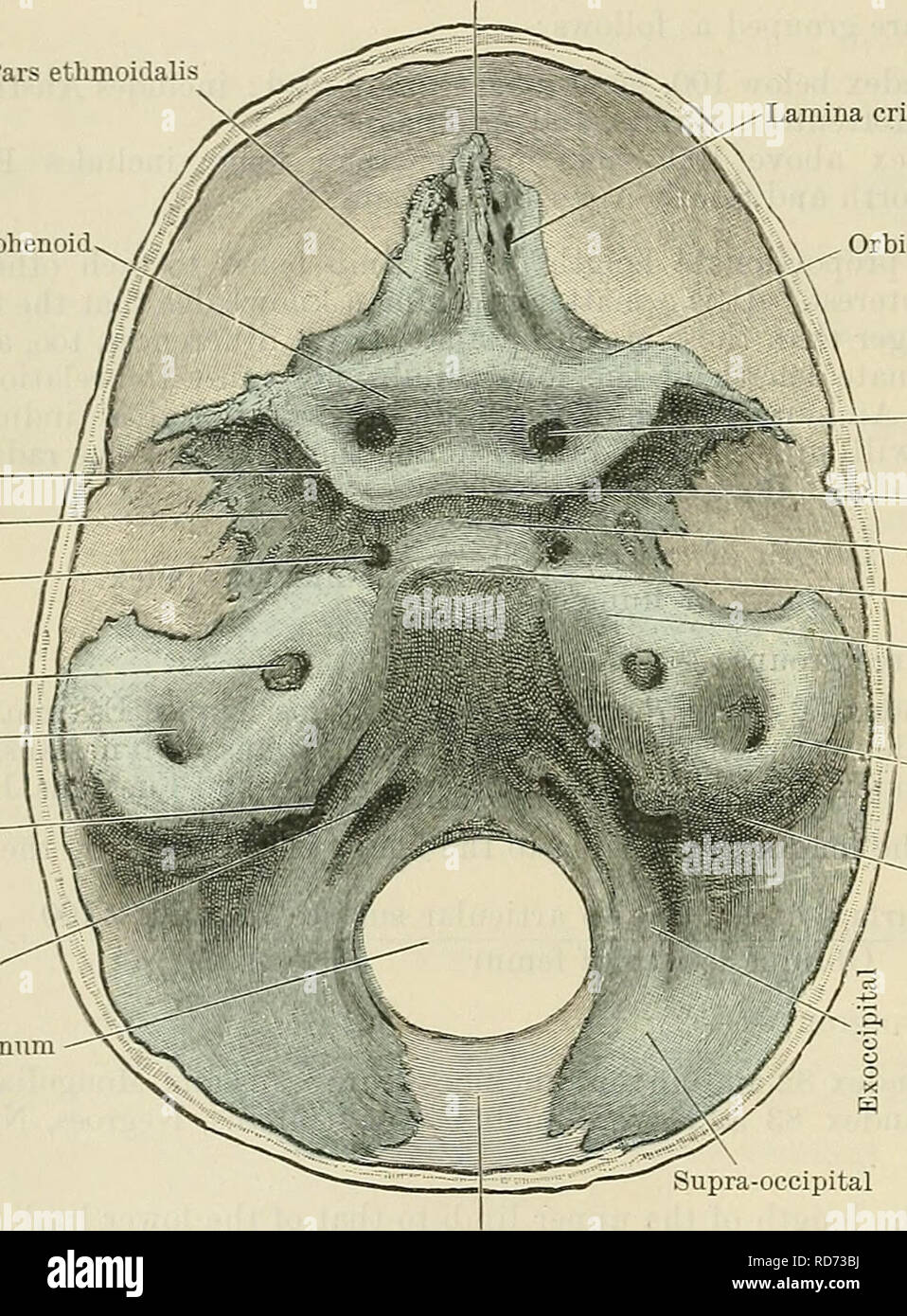
. Cunningham's Text-book of anatomy. Anatomy. 290 OSTEOLOGY. APPENDIX E. DEVELOPMENT OF THE CHONDRO-CRANIUM AND MORPHOLOGY OF THE SKULL. As has been already stated, the chorda dorsalis or notochord extends forwards to a point immediately beneath the anterior end of the mid-brain. In front of this the head takes a bend so that the large fore-brain overlaps the anterior extremity of the notochord. At this stage of development the cerebral vesicles are enclosed in a membranous covering derived from the mesen- Crista Galli Pars ethmoidals Lamina cribrosa Orbito-sphenoid Superior orbital fissure 77J/ Alisphenoid jj Carotid canal la Meatus acustieus li If internus Subareuate fossa Jugular foramen Canalis hypoglossi Foramen magnum. Orbital portion of orbito-sphenoid Optic foramen Tuberculum sell* 1 (Olivary process) Sella turcica — Dorsum sellse ^~p Pars petrosa Superior semicircular canal Pars mastoidea Supra-occipital Occipital fontanelle Fig. 282.—View of the Chondro- Cranium of a Human Fcetus 5 cm. in length from Vertex to Coccyx (about the middle of the third month) ; the cartilage is coloured blue. The line to the right of the drawing shows the actual size. chyme surrounding the notochord ; this differentiated mesodermal layer is called the primordial membranous cranium. Prom it the meninges which invest the brain are derived. In lower vertebrates this membranous capsule becomes converted into a thick-walled cartilaginous envelope, the primordial cartilaginous cranium. In mammals, however, only the basal part of this capsule becomes chondrified, the roof and part of the sides remaining membranous. In considering the chondrification of the skull in mammals, it must be noted that part only of the base is traversed by the notochord, viz., that portion which extends from the foramen magnum to the dorsum sella? of the sphenoid. It is, therefore, conveniently divided into two parts—one posterior, surrounding the notochord, and hence called chordal, and one in fro