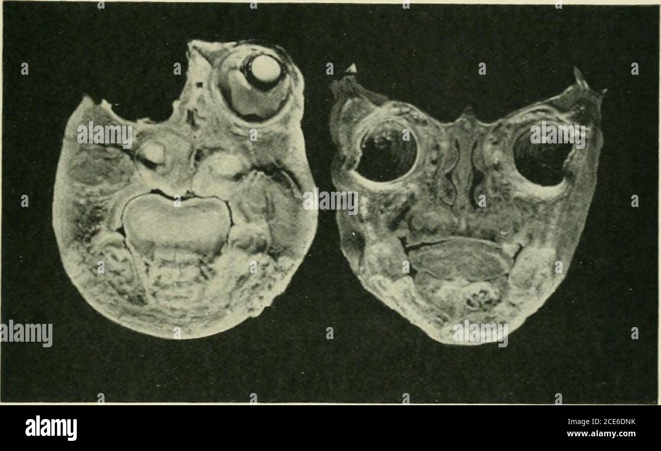
. Journal of anatomy . -MA tS Dl OL e. Fig, 1.—Coronal section of the face of a Cyclopean monster (Mus.Anat. Cant, specimen HC ), drawn with Edingers pro-jection apparatus. the septum is reduced to the merest downward projection. No retinalelements have been detected {cf. condition described in cyclopia by Leonowa,Archiv filr Psychiatrie, Band xxxviii.). The maxillary processes havefused to form the hard palate. The posterior orifice of the conjoined nasalfossae is reduced to a mere pinhole aperture. Cyclopean F(l4iis with Hernia Encephali 59 II. Eyeball and Retina. The eyeball presents no mar
Image details
Contributor:
Reading Room 2020 / Alamy Stock PhotoImage ID:
2CE6DNKFile size:
7.2 MB (236.6 KB Compressed download)Releases:
Model - no | Property - noDo I need a release?Dimensions:
2019 x 1238 px | 34.2 x 21 cm | 13.5 x 8.3 inches | 150dpiMore information:
This image is a public domain image, which means either that copyright has expired in the image or the copyright holder has waived their copyright. Alamy charges you a fee for access to the high resolution copy of the image.
This image could have imperfections as it’s either historical or reportage.
. Journal of anatomy . -MA tS Dl OL e. Fig, 1.—Coronal section of the face of a Cyclopean monster (Mus.Anat. Cant, specimen HC ), drawn with Edingers pro-jection apparatus. the septum is reduced to the merest downward projection. No retinalelements have been detected {cf. condition described in cyclopia by Leonowa, Archiv filr Psychiatrie, Band xxxviii.). The maxillary processes havefused to form the hard palate. The posterior orifice of the conjoined nasalfossae is reduced to a mere pinhole aperture. Cyclopean F(l4iis with Hernia Encephali 59 II. Eyeball and Retina. The eyeball presents no marked deviation fioni normal conditions.But, if the eyeball of a full-time fietus is employed for purposes of control, the following ditterences appear:— («) The optic nerve cells are more scanty and are smaller in theCyclopean specimen. {h) The whole retina is shallower. This, of course, may be due to theearlier age of the specimen, but it is noteworthy that the main dlHcrences. Fig. 2.—Photographs of coronal sections of a Cyclopean monster ( HC ), and ...f anormal seventh mouth fcetus used as a control. Of the first specimen, theright eye has been removed for histological examination, appear in the layer of rods and cones and the strata which would corre-spond to the molecular layers in the normal eye. These layers are muchbetter developed in the contiol. On the other hand, the inner nuclearlayer is, relatively speaking, as distinct as it is in the control. III. Cextkal Nervous System. The cerebral mass was so divided as to provide a block from the leftside for sagittal sections and another from the right side for coronal sections.There is a single ventricular cavity. The brain substance contains anexcess of nuclei, although the stratitication, of cells at present small, inanticipation of the future cortical lamination, has already connuenced. Itis possible to di.stinguish an outer molecular layer, two granular layers, 60 Mr F. W. Watkyn-Thoinas and an inf