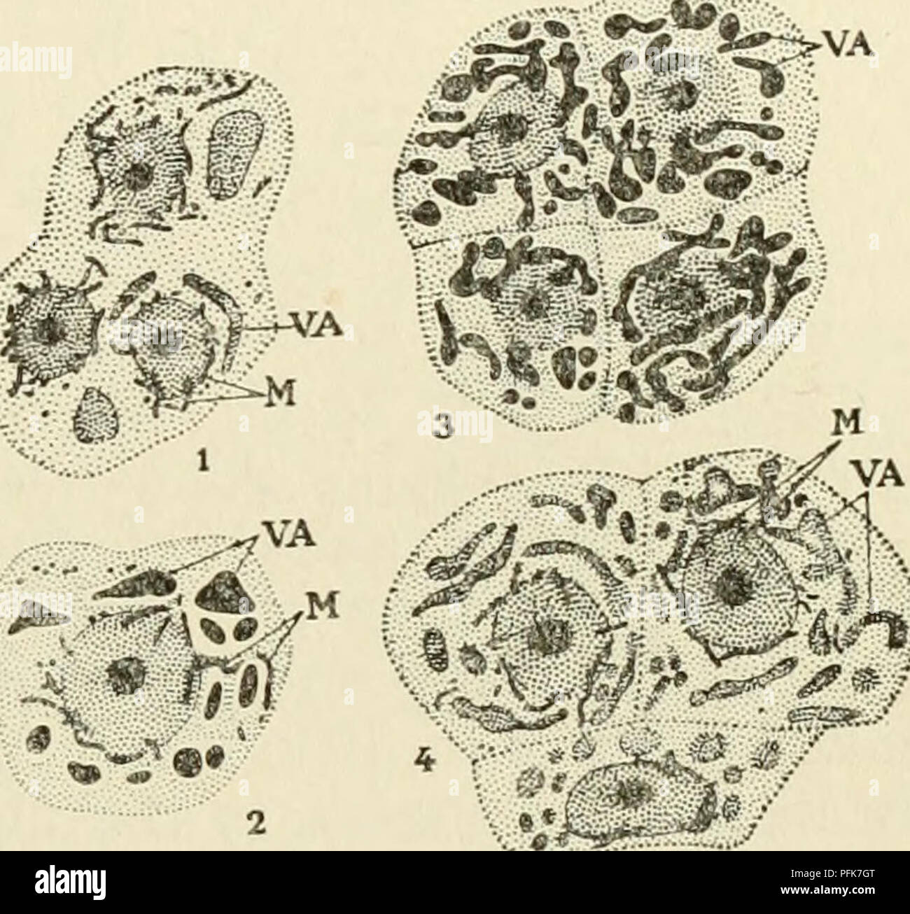
. The cytoplasm of the plant cell. Plant cells and tissues; Protoplasm. Chapter XIV 147 — The Vacuolar System pigment in solution. By reason of the great resemblance between the initial shapes in which anthoeyanin first appears and the shapes of the chondrioconts, we were led to think, originally, that this pigment arose in the elements of the chondriome which then be- came transformed into vacuoles. This interpretation was founded also on the fact that these shapes were preserved by mitochondrial techniques. With the method of Regaud, for example, we ob- tained at that time, both typical chon
Image details
Contributor:
Central Historic Books / Alamy Stock PhotoImage ID:
PFK7GTFile size:
7.1 MB (378.6 KB Compressed download)Releases:
Model - no | Property - noDo I need a release?Dimensions:
1634 x 1529 px | 27.7 x 25.9 cm | 10.9 x 10.2 inches | 150dpiMore information:
This image is a public domain image, which means either that copyright has expired in the image or the copyright holder has waived their copyright. Alamy charges you a fee for access to the high resolution copy of the image.
This image could have imperfections as it’s either historical or reportage.
. The cytoplasm of the plant cell. Plant cells and tissues; Protoplasm. Chapter XIV 147 — The Vacuolar System pigment in solution. By reason of the great resemblance between the initial shapes in which anthoeyanin first appears and the shapes of the chondrioconts, we were led to think, originally, that this pigment arose in the elements of the chondriome which then be- came transformed into vacuoles. This interpretation was founded also on the fact that these shapes were preserved by mitochondrial techniques. With the method of Regaud, for example, we ob- tained at that time, both typical chondriosomes and elements of the same form as the chondriosomes, but a little larger, stained in the same way, but for which the staining was less stable and which, if destaining was prolonged, lost all the dye and took a yellow color. This color we attributed to the action of the potas- sium bichromate on the anthoey- anin. Now, between the typical chondriosomes and these elements which resemble them, there seemed to exist all intermediate stages. We thought at that time, therefore, that the larger elements, to which the potassium bichromate gives a yellow color, corresponded to chondriosomes impregnated with anthoeyanin. Our interpretation was a natural one at the moment, for the chon- driome was not yet well known, the origin of the vacuoles was not understood, and it was thought that most of the pigments of ani- mal cells were of mitochondrial origin. This observation was immedi- ately investigated by a certain number of workers among whom some agreed with our interpreta- tions (MoREAU, MiRANDE) and others contested them. Among the latter Arthur Meyer thought, but without having proved it, that the chondriosome-like elements which mark the beginning of the formation of anthoeyanin, represent filamentous vacuoles. Low- SCHIN, on the other hand, expressed the opinion that these fig- ures correspond merely to anthoeyanin itself. According to him the anthoeyanin is deposited in