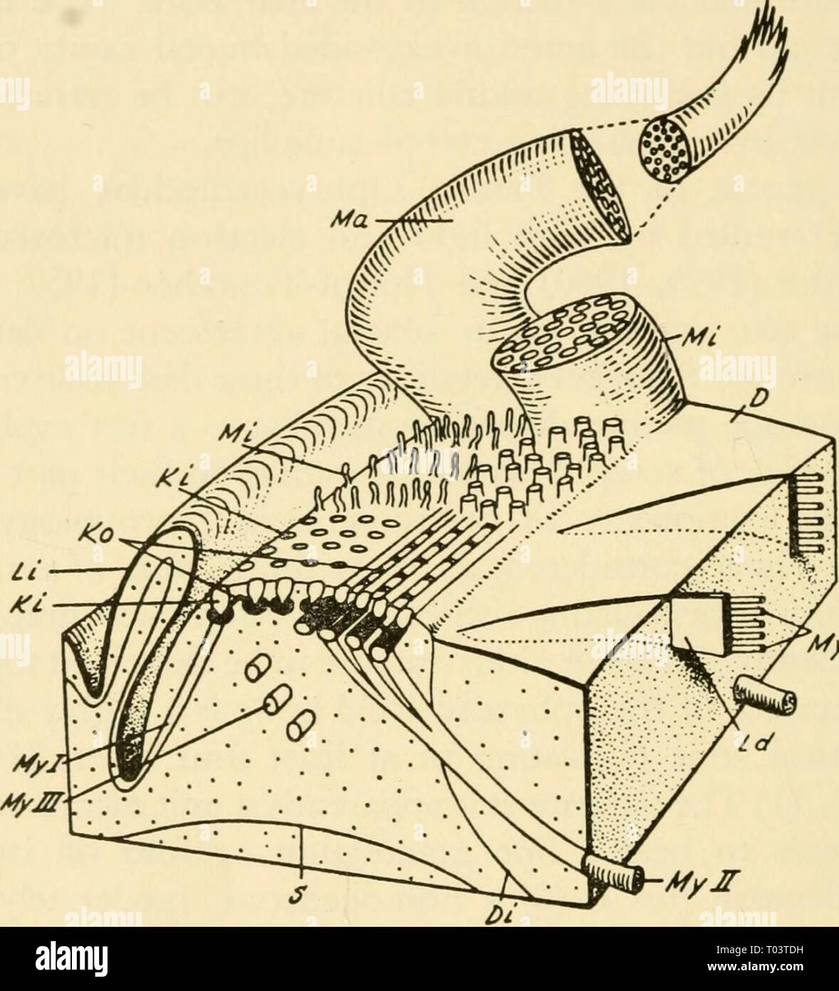
Electron-microscopic structure of protozoa . electronmicrosco00pite Year: 1963 208 ELECTRON-MICROSCOPIC STRUCTURE OF PROTOZOA The adoral ciliature consists of a long spiral band in which cilia occur in short oblique rows, each row arising from a basal rod visible in the light microscope (Text-fig. 16). Electron micrographs of sections through the level of the kinetosomes show that the rows are uniformly aligned, and not separated into groups corresponding to discrete membranelles. Each kineto- Mymr Text-figure 16. Diagrammatic reconstruction of part of the adoral ciliary zone of an ophryos
Image details
Contributor:
Bookend / Alamy Stock PhotoImage ID:
T03TDHFile size:
5.7 MB (212.9 KB Compressed download)Releases:
Model - no | Property - noDo I need a release?Dimensions:
1346 x 1485 px | 22.8 x 25.1 cm | 9 x 9.9 inches | 150dpiMore information:
This image is a public domain image, which means either that copyright has expired in the image or the copyright holder has waived their copyright. Alamy charges you a fee for access to the high resolution copy of the image.
This image could have imperfections as it’s either historical or reportage.
Electron-microscopic structure of protozoa . electronmicrosco00pite Year: 1963 208 ELECTRON-MICROSCOPIC STRUCTURE OF PROTOZOA The adoral ciliature consists of a long spiral band in which cilia occur in short oblique rows, each row arising from a basal rod visible in the light microscope (Text-fig. 16). Electron micrographs of sections through the level of the kinetosomes show that the rows are uniformly aligned, and not separated into groups corresponding to discrete membranelles. Each kineto- Mymr Text-figure 16. Diagrammatic reconstruction of part of the adoral ciliary zone of an ophryoscolecid ciliate. Two syncilia are shown at the rear, labeled Ma and Mi. In front of these are shown, on the right, the stubs of cilia making up another syncilium and, on the left, microvilli labeled Mi, that protrude from the cell membrane between cilia. In front of this are the apices of kineto- somes (Ki) on the left and the outlines of basal rods on the right. Noirot-Timothee has shown that in at least some species the basal rods pass obliquely across the band rather than longitudinally as shown here. Commissural fibers (Ko) are shown connecting the kinetosomes laterally. On the cut faces of the stereogram are seen various fibrous structures considered by Bretschneider to be myonemes. My I, II, and III are retrociliary fibers passing in different directions. My IV and Ld together are the fibrillar sheets subtending the peristomial disc (D). From Bretschneider, 1960.