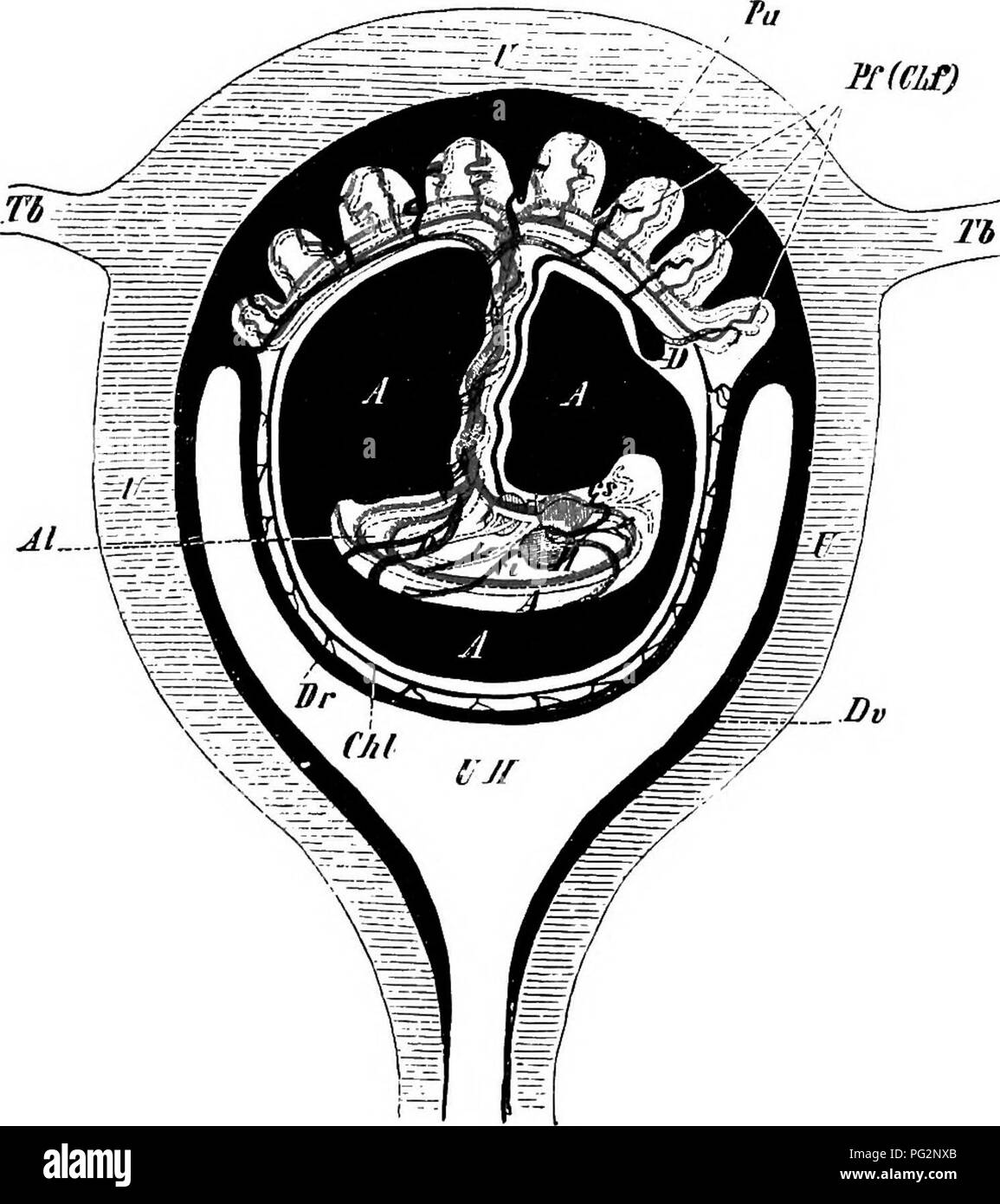
. Elements of the comparative anatomy of vertebrates. Anatomy, Comparative. INTRODUCTION 11 Ulaierally symmetrical. The neural tube, or cerebro-spinal cavity, enclosed by the skull and vertebral arches, contains the central ner- TTOUS system {hrain and spinal cord); the visceral tube (ccelome, p. 8, Fig. 7) encloses the viscera (alimentary canal, urinogenital -organs, &c.),and its muscular walls may be strengthened by a series l'f(CLf). Fig. 9.—Diagrammatic Section through the Human Gravid Uteris. U, uterus ; Th, Tb, Fallopian tubes ; UH, uterine cavity ; Dv, deeitlua vera, which at Pu pas
Image details
Contributor:
Central Historic Books / Alamy Stock PhotoImage ID:
PG2NXBFile size:
7.2 MB (327.7 KB Compressed download)Releases:
Model - no | Property - noDo I need a release?Dimensions:
1491 x 1677 px | 25.2 x 28.4 cm | 9.9 x 11.2 inches | 150dpiMore information:
This image is a public domain image, which means either that copyright has expired in the image or the copyright holder has waived their copyright. Alamy charges you a fee for access to the high resolution copy of the image.
This image could have imperfections as it’s either historical or reportage.
. Elements of the comparative anatomy of vertebrates. Anatomy, Comparative. INTRODUCTION 11 Ulaierally symmetrical. The neural tube, or cerebro-spinal cavity, enclosed by the skull and vertebral arches, contains the central ner- TTOUS system {hrain and spinal cord); the visceral tube (ccelome, p. 8, Fig. 7) encloses the viscera (alimentary canal, urinogenital -organs, &c.), and its muscular walls may be strengthened by a series l'f(CLf). Fig. 9.—Diagrammatic Section through the Human Gravid Uteris. U, uterus ; Th, Tb, Fallopian tubes ; UH, uterine cavity ; Dv, deeitlua vera, which at Pu passes into the uterine portion of the placenta ; Dr, decidua reflexa ; Pf, fcutal portion of the placenta (chorion frondosum, Chf); Chi, chorion liKve lAtA, the cavity of the amnion filled with iluid : in the interior of the amnion is seen the embryo suspended by the twisted umbilical cord; H, neart; A, aorta ; cs, precaval, ci, postcaval, and p, portal vein ; Al, allantoic (umbilical) arteries; t, the liver, perforated by the umbilical vein; D, the remains of the yolk-sac (umbilical vesicle). of ribs, articulating dorsally with the vertebral column. Certain of the ribs may reach the mid-ventral line and come into connec- tion with a breast-bone or sternum, and thus form complete rings •or hoops around the visceral tube. The anterior ends of the central nervous system (brain) and ali- mentary tract enter into close relations with the outer world, the. Please note that these images are extracted from scanned page images that may have been digitally enhanced for readability - coloration and appearance of these illustrations may not perfectly resemble the original work.. Wiedersheim, Robert, 1848-1923; Parker, William Newton, 1857-1923. London, Macmillan