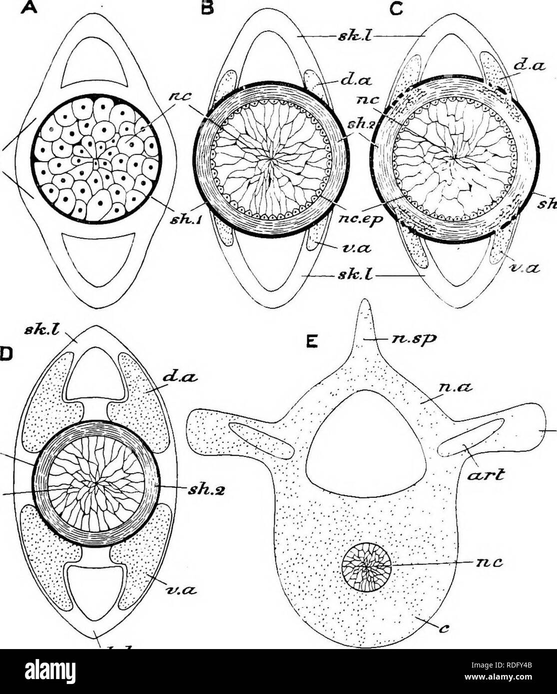
. Elements of the comparative anatomy of vertebrates. Anatomy, Comparative. VEBTEBRAL COLU^IX 35 sJc.h sTi.i sh.i. tr-.p Fig. 22.- A.- sJc-.l -Diagrams illdsteatixg the Development of the Notochokdal Sheaths and Vertebral Column. B. C. -Early stage, showing notoehordal cells (hc) and primary sheath (nh^), as well as the mesoblastio skeletogenoiis layer {sk.l). -Later stage, in which the central notoehordal cells (nr) have become vacuolated and the peripheral cells have given ri.se to the " notoehordal epithe- limn" (rec. ep.) from which the fibrillar secondary sheath (sW) is derived
Image details
Contributor:
The Book Worm / Alamy Stock PhotoImage ID:
RDFY4BFile size:
7.1 MB (395.3 KB Compressed download)Releases:
Model - no | Property - noDo I need a release?Dimensions:
1464 x 1706 px | 24.8 x 28.9 cm | 9.8 x 11.4 inches | 150dpiMore information:
This image is a public domain image, which means either that copyright has expired in the image or the copyright holder has waived their copyright. Alamy charges you a fee for access to the high resolution copy of the image.
This image could have imperfections as it’s either historical or reportage.
. Elements of the comparative anatomy of vertebrates. Anatomy, Comparative. VEBTEBRAL COLU^IX 35 sJc.h sTi.i sh.i. tr-.p Fig. 22.- A.- sJc-.l -Diagrams illdsteatixg the Development of the Notochokdal Sheaths and Vertebral Column. B. C. -Early stage, showing notoehordal cells (hc) and primary sheath (nh^), as well as the mesoblastio skeletogenoiis layer {sk.l). -Later stage, in which the central notoehordal cells (nr) have become vacuolated and the peripheral cells have given ri.se to the " notoehordal epithe- limn" (rec. ep.) from which the fibrillar secondary sheath (sW) is derived : paired dorsal and ventral cartilages {d.a, i.a) have arisen in the skeletogenoiis layer. Cartilage cells have passed through the primary sheath, and are invading the secondary sheath (Cartilaginous Ganoids, Holocephali, Dipnoi, Elasmo- branohii : in the last named chorda-centra are thus formed). D.—The cartilages are growing round the notochord, outside its sheaths, which gradually become reduced : thus arch-centra are formed (Bony Ganoids, Teleostei, Amphibia, Amniota). A—D represent the caudal region. E.—A later stage in the development of a pre-caudal vertebra. The notochord (nc) has become constricted, and the cartilages have united into a single mass and have given rise to a centrum (c), neural arch (jt.re), neural spine {n. sp), transverse processes (tr.p) and articular processes (art); D 2. Please note that these images are extracted from scanned page images that may have been digitally enhanced for readability - coloration and appearance of these illustrations may not perfectly resemble the original work.. Wiedersheim, Robert, 1848-1923; Parker, William Newton, 1857-1923. London, Macmillan