Illustration showing the structure of the retina as an insert to the larger eye structure. From top to bottom: optic nerve fiber (reddish brown strip at top), ganglion cells (in brown), amacrine cells (in dark blue), bipolar cells (in brownish-orange), ho
RMID:Image ID:G1564N
Image details
Contributor:
Science History Images / Alamy Stock PhotoImage ID:
G1564NFile size:
38.4 MB (598.2 KB Compressed download)Releases:
Model - no | Property - noDo I need a release?Dimensions:
4108 x 3269 px | 34.8 x 27.7 cm | 13.7 x 10.9 inches | 300dpiPhotographer:
Spencer SuttonMore information:
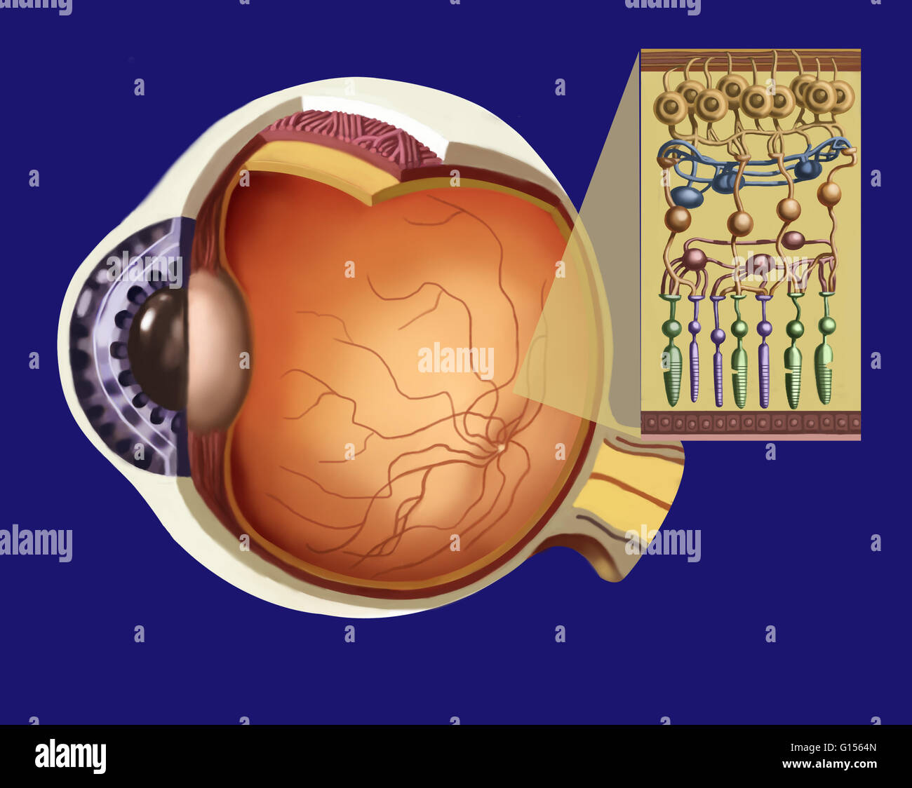
Illustration showing the structure of the retina as an insert to the larger eye structure. From top to bottom: optic nerve fiber (reddish brown strip at top), ganglion cells (in brown), amacrine cells (in dark blue), bipolar cells (in brownish-orange), horizontal cells (in dark pink), Muller glial cell (in beige), photoreceptors: rods (in purple), cones (in green), retina pigment epithelium (bottom strip) with the pigmentary cells (inside bottom strip), Bruch membrane (at very bottom in pink).