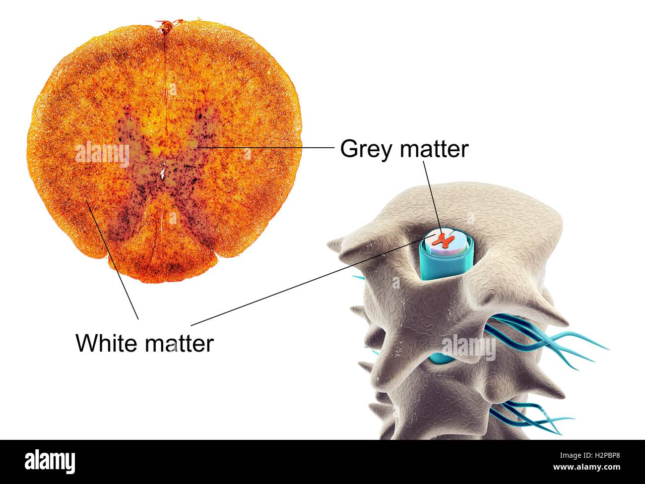
Spinal cord, cross-section. Light micrograph and computer illustration of a spinal cord. At right it is seen inside the vertebrae (bones). The section at left shows the white and grey matter with dorsal and ventral horns.
RFID:Image ID:H2PBP8
Image details
Contributor:
Science Photo Library / Alamy Stock PhotoImage ID:
H2PBP8File size:
84.9 MB (3.1 MB Compressed download)Releases:
Model - no | Property - noDo I need a release?Dimensions:
6596 x 4500 px | 55.8 x 38.1 cm | 22 x 15 inches | 300dpiDate taken:
26 September 2016Photographer:
KATERYNA KON/SCIENCE PHOTO LIBRARY