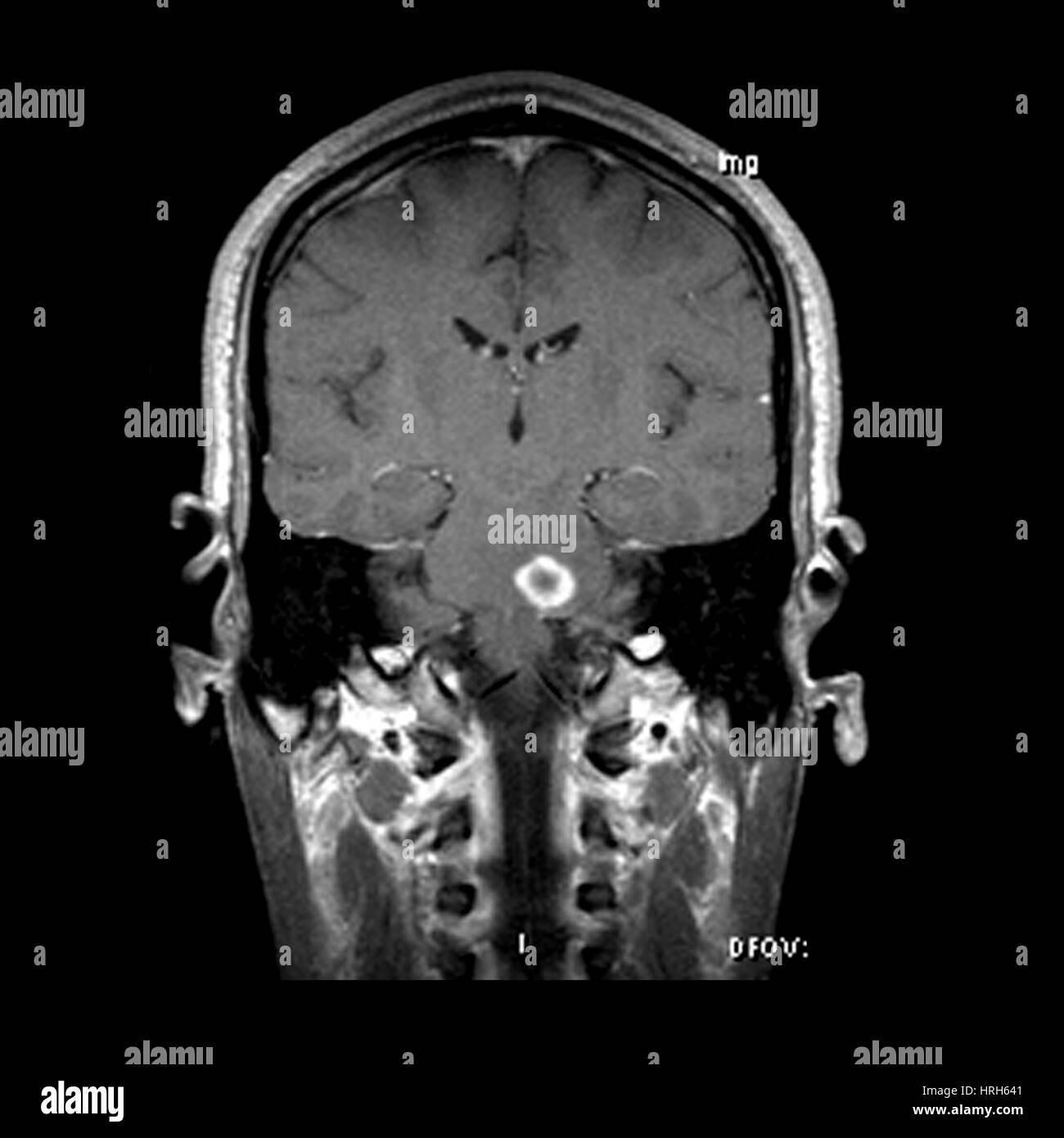
MRI of Acute MS
RMID:Image ID:HRH641
Image details
Contributor:
Science History Images / Alamy Stock PhotoImage ID:
HRH641File size:
37.1 MB (424.3 KB Compressed download)Releases:
Model - no | Property - noDo I need a release?Dimensions:
3600 x 3600 px | 30.5 x 30.5 cm | 12 x 12 inches | 300dpiPhotographer:
Medical Body ScansMore information:
This image could have imperfections as it’s either historical or reportage.
This coronal (frontal) contrast enhanced MRI image of the brain through the level of the brain stem reveals a prominent ring enhancing lesion in the left side of the pons. T2 weighted images demonstrated extensive, mass-like brain stem edema and swelling. This represents a form of acute MS that can stimulate a tumor therefore it is referred to as tumefactive.