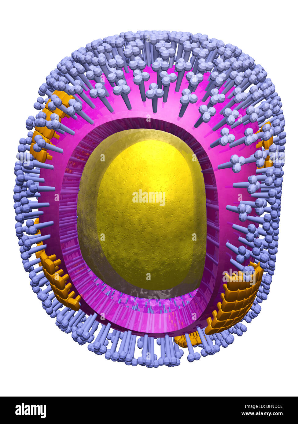
Three-dimensional computer-generated model of the structure of the H1N1 swine influenza virus particle.
Image details
Contributor:
Scott Camazine / Alamy Stock PhotoImage ID:
BFNDCEFile size:
58 MB (1.7 MB Compressed download)Releases:
Model - no | Property - noDo I need a release?Dimensions:
3900 x 5200 px | 33 x 44 cm | 13 x 17.3 inches | 300dpiDate taken:
2009More information:
Three-dimensional computer-generated model of the structure of the H1N1 swine influenza virus particle. The surface of the virus is studded with two types of subunits, hemagglutinins (HA) and neuraminidases (NA). HA are glycoproteins that mediate binding of the virus to target cells and entry of the viral genome into the target cell, while NA is involved in the release of progeny virus from infected cells, by cleaving sugars that bind the mature viral particles. In this model, the hemagglutinins are the represented by the large number of structures, each with 3 spherical knobs which cover the surface of the virus particle (light blue) . The neuraminidase subunits are seen as smaller patches of structures that do not stick out as far from the surface (gold). In the 2009 flu pandemic, the virus isolated from patients in the United States was found to be made up of genetic elements from four different flu viruses – North American swine influenza, North American avian influenza, human influenza, and swine influenza virus typically found in Asia and Europe