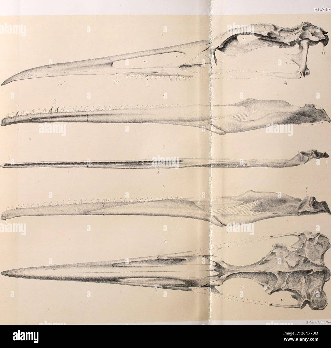
. Report of the geological exploration of the fortieth parallel . Fr--:- PLATE I * F EergK, del. Cnsand, litii NBwHavo; HESPERORNIS REGALIS, Mars}i. PL^TE II. PL^TE II. ODONTOR^TITHES. Teeth ajtid Skull of Hespekobnis regalis Marsh. All Figures Natural Size, except 2 and 3. Page. Fig. 1.—Inferior 6urf;ce of iircmaxillaiy and maxillary bones, 8 t —Tooth iu maxillary. Fig. 2.—Tooth; from lower jaw, lateral view, seen from the left; magnified eight diameters,.. 13 Fig. 3.—Tooth; from maxillary, lateral view, seen from the right; magnified eight diameters,. 13a —Young tooth, showing mode of devel
Image details
Contributor:
Reading Room 2020 / Alamy Stock PhotoImage ID:
2CNX7DMFile size:
7.1 MB (280.6 KB Compressed download)Releases:
Model - no | Property - noDo I need a release?Dimensions:
1595 x 1566 px | 27 x 26.5 cm | 10.6 x 10.4 inches | 150dpiMore information:
This image could have imperfections as it’s either historical or reportage.
. Report of the geological exploration of the fortieth parallel . Fr--:- PLATE I * F EergK, del. Cnsand, litii NBwHavo; HESPERORNIS REGALIS, Mars}i. PL^TE II. PL^TE II. ODONTOR^TITHES. Teeth ajtid Skull of Hespekobnis regalis Marsh. All Figures Natural Size, except 2 and 3. Page. Fig. 1.—Inferior 6urf;ce of iircmaxillaiy and maxillary bones, 8 t —Tooth iu maxillary. Fig. 2.—Tooth; from lower jaw, lateral view, seen from the left; magnified eight diameters, .. 13 Fig. 3.—Tooth; from maxillary, lateral view, seen from the right; magnified eight diameters, . 13a —Young tooth, showing mode of development. Fig. 4.—Base of skull; posterior view. Top of cranium broken away, - 6 bp —Basi-pterygoid process.fm —Foramen magnum. Fig. 5.—Base of skull; seen from below, . - 6 Fig. 6.—Left quadrate hone ; inner view, - — - 6 6a —Superior view, showing undivided articulation.66 —Posterior view.6c —Inferior view. Fig. V.—^Posterior part of right pterygoid bone ; inferior view, C la —Posterior view, showing concave articulation for quadrate.lb —Superior view. Fig.