English: Illustration from Report on the Radiolaria collected by H.M.S. Challenger during the years 1873-1876. Part III. Original description follows: Plate 100. Tuscarorida. Diam. Fig. 1. Tuscarora bisternaria, John Murray, × 30 View from the dorsal side. Fig. 1a. View from the mouth pole × 25 Fig. 2. Tuscarora murrayi, n. sp., × 30 View from the dorsal side. The central capsule (in the aboral half), and the phæodium (in the middle of the shell-cavity) are visible. A fine network of pseudopodia pierces the calymma, which fills up the shell-cavity. Fig. 3. Tuscarora wyvillei, n. sp., ×
Image details
Contributor:
History and Art Collection / Alamy Stock PhotoImage ID:
P7B31BFile size:
14.3 MB (533.2 KB Compressed download)Releases:
Model - no | Property - noDo I need a release?Dimensions:
2000 x 2500 px | 16.9 x 21.2 cm | 6.7 x 8.3 inches | 300dpiMore information:
This image is a public domain image, which means either that copyright has expired in the image or the copyright holder has waived their copyright. Alamy charges you a fee for access to the high resolution copy of the image.
This image could have imperfections as it’s either historical or reportage.
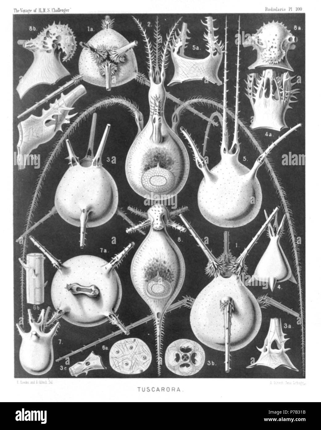
English: Illustration from Report on the Radiolaria collected by H.M.S. Challenger during the years 1873-1876. Part III. Original description follows: Plate 100. Tuscarorida. Diam. Fig. 1. Tuscarora bisternaria, John Murray, × 30 View from the dorsal side. Fig. 1a. View from the mouth pole × 25 Fig. 2. Tuscarora murrayi, n. sp., × 30 View from the dorsal side. The central capsule (in the aboral half), and the phæodium (in the middle of the shell-cavity) are visible. A fine network of pseudopodia pierces the calymma, which fills up the shell-cavity. Fig. 3. Tuscarora wyvillei, n. sp., × 30 View from the dorsal side. Fig. 3a. Base of a tooth, × 100 Fig. 3b. Transverse section through the base of a tooth. Fig. 3c. Base of a foot. Fig. 4. Tuscarora tetrahedra, John Murray, × 15 View from the dorsal side. Fig. 4a. Mouth with the three teeth, × 50 Fig. 5. Tuscarora tubulosa, John Murray, × 40 View from the ventral side. Fig. 5a. Mouth with the two teeth, × 100 Fig. 5b. Basal part of a single tooth, × 150 Fig. 6. Tuscarora porcellana, John Murray, × 600 Fig. 6a. A piece of the shell, with five pores. Fig. 6b. A piece of a tooth, with the internal axial rod and its transverse branches. Fig. 7. Tuscarusa medusa, n. sp., × 25 View from the side. Fig. 7a. View from the mouth, × 50 Fig. 8. Tuscaridium lithornithium, n. sp., × 20 View from the ventral side. Central capsule and calymma as in fig. 2. Fig. 8a. Peristome from the ventral side. Fig. 8b. Peristome from the right side. . 1887 66 Radiolaria (Challenger) Plate 100