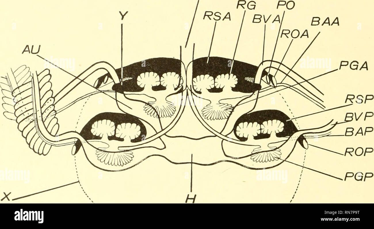
. The anatomy of Nautilus pompilius. Nautilus. 164 MEMOIRS OF THE NATIONAL ACADEMY OF SCIENCES. The other two reiiiil sacs, the i)osterior .saes. lie at the sides of the Ixxly. widely sei)arate(l from each other. A little posterior to the inner sacs, they are eon.siderahly outside them, to close the ventral sides of the sii(>ll muscles. The dorsal wall of each sac is formed by the inner wall of the mantle fold, and is that portion of the inner wall of the mantle fold lying between the anterior ri'iial pores and the base of the posterior gill. The posterior and anterior walls of the sac are
Image details
Contributor:
Library Book Collection / Alamy Stock PhotoImage ID:
RN7P9TFile size:
7.1 MB (265.8 KB Compressed download)Releases:
Model - no | Property - noDo I need a release?Dimensions:
2140 x 1167 px | 36.2 x 19.8 cm | 14.3 x 7.8 inches | 150dpiMore information:
This image is a public domain image, which means either that copyright has expired in the image or the copyright holder has waived their copyright. Alamy charges you a fee for access to the high resolution copy of the image.
This image could have imperfections as it’s either historical or reportage.
. The anatomy of Nautilus pompilius. Nautilus. 164 MEMOIRS OF THE NATIONAL ACADEMY OF SCIENCES. The other two reiiiil sacs, the i)osterior .saes. lie at the sides of the Ixxly. widely sei)arate(l from each other. A little posterior to the inner sacs, they are eon.siderahly outside them, to close the ventral sides of the sii(>ll muscles. The dorsal wall of each sac is formed by the inner wall of the mantle fold, and is that portion of the inner wall of the mantle fold lying between the anterior ri'iial pores and the base of the posterior gill. The posterior and anterior walls of the sac are formed by septa extending backward and downward from tlu' inner wall of the mantle. They unite around the lobular appendages to form a closed sac, from the posterior edge of which a thin ligament extends back- ward and somewhat inward, attached along one edge to the visceral body wall. (Fig. 3»5, L.) The outer end of the sac is narrowed to form a canal which, running in the substance of the mantle vc RG PO RSA I BVAJ BAA ROA. TEXT-FRi y -DIACiRAM OF THK RENAL SACS AND NEIGHBORING OliGANS OF NAUTILUS, AS VIEWED FROM THE DORSAL SIDE. R(t, anterior renal appendage. ROA, antericif ivnal i«iie. ROP, posterior renal pore. RSA, anterior renal sac. RSI', posterior renal sac. VC, vena cava. X, outline of pericanlial divitiion of cceloiu. Y, cul de sac of anterior renal sac. AU, anricnlar expansion of left anterior branchial vein. HA., anterior liranchial artery. B.P, posterior branchial artery. I?V., anterior liranihial vein. BVP, posterior liranchial vein. H, heart. PGA, anterior pericardial appendage. PGP, posterior pericardial appendage. PO, pericardial pore. close to the ventral surface of the shell muscle outside and ventral to the liranchial artery, opens to the exterior through one of the posterior renal pores. (Fig. 3, RP.) The posterior branchial arteries run through th(> posterior walls of the outer renal .sacs. (Text-tig. tt, BAP.) Between the inner and outer renal sa