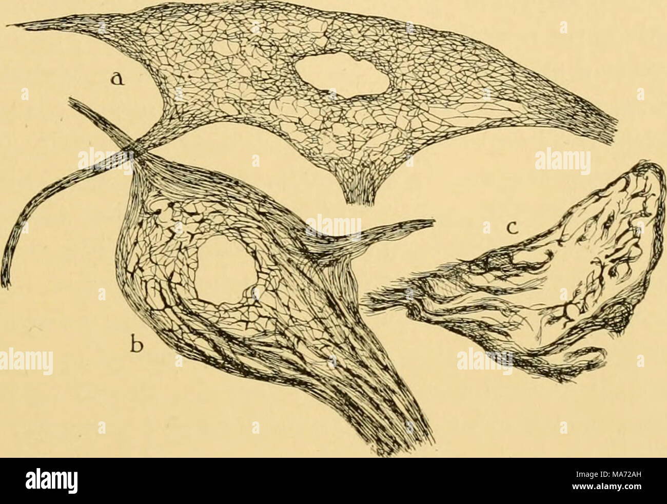
. The effects of inanition and malnutrition upon growth and structure . Fig. 62.—Nerve cells from the ventral horn of the spinal cord in the dog, showing the variable changes in the neurofibrillae by Donaggio's silver method after a loss of about 50 per cent in body weight from total inanition, a, a cell in which the neurofibrillae have retained nearly normal structure; b, cell showing some vacuolation in the perinuclear region; c, cell with disorganized neurofibrillae crowded into bundles between the numerous large cytoplasmic vacuoles. (Riva '06.) literature on these effects of inanition upo
Image details
Contributor:
The Bookworm Collection / Alamy Stock PhotoImage ID:
MA72AHFile size:
14.3 MB (545.2 KB Compressed download)Releases:
Model - no | Property - noDo I need a release?Dimensions:
2699 x 1852 px | 22.9 x 15.7 cm | 9 x 6.2 inches | 300dpiMore information:
This image is a public domain image, which means either that copyright has expired in the image or the copyright holder has waived their copyright. Alamy charges you a fee for access to the high resolution copy of the image.
This image could have imperfections as it’s either historical or reportage.
. The effects of inanition and malnutrition upon growth and structure . Fig. 62.—Nerve cells from the ventral horn of the spinal cord in the dog, showing the variable changes in the neurofibrillae by Donaggio's silver method after a loss of about 50 per cent in body weight from total inanition, a, a cell in which the neurofibrillae have retained nearly normal structure; b, cell showing some vacuolation in the perinuclear region; c, cell with disorganized neurofibrillae crowded into bundles between the numerous large cytoplasmic vacuoles. (Riva '06.) literature on these effects of inanition upon the nerve cells in general is fully reviewed by Marinesco ('09). In Necturus maculatiis starved 4 months the spinal cord was found nearly normal by Smallwood and Rogers ('n). After 16 months, however, the spinal cord appears greatly reduced in size, with atrophy especially of the gray sub- stance. Changes found in the spinal ganglia will be mentioned in the next chapter. In the nerve cells of the spinal cord in Triton cristatus fasting 1-6 months, Frankenberger ('17) described nuclear changes resembling pycnosis. In starved albino rats (on water only), Sundwall ('17) noted marked conges- tion and edema of the spinal cord; anterior horn cells swollen, vacuolated and chromatolytic, with vesicular or pycnotic nuclei. There is diffuse degeneration of the various nerve fiber tracts, especially in the posterior columns. Changes during Hibernation.—Since hibernation represents a special type of inanition, the corresponding changes in the nerve cells of the spinal cord may be separately considered. (The observations on the brain cells during hiberna- tion by Querton '98, Legge '99, BaronciniandBeretta'ooand Marinesco'05 were mentioned in Chapter X.) First as to the Nissl substance, no appreciable change