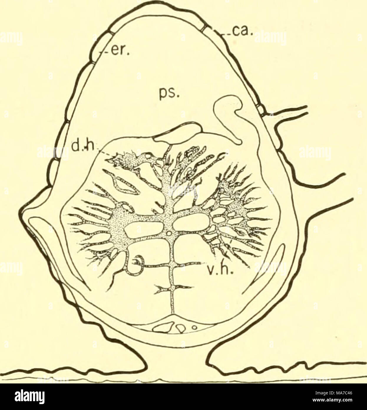
. The elasmobranch fishes . A B Fig. 217. Transverse sections of the spinal cord. (From Sterzi.) A. Acanthias vulgaris. B. Baja clavata. ca., calcification; d.li., dorsal horn; d.r., dorsal root; er., endorachis; nc, neurocoele; pm., paracentral mass; ps., perimeningeal space; v.h., ventral horn. sixth nerve and the grey matter of the formatio-reticularis {f.r., fig. 216) is occupied in the cord by the ventral horn {v.h., fig. 217); while the general cutaneous nucleus of the medulla (g.c.n.) gives place to the dorsal horn of the cord (d.h., fig. 217); and the lobes of the vagus and the viscero
Image details
Contributor:
The Bookworm Collection / Alamy Stock PhotoImage ID:
MA7C46File size:
14.3 MB (335.8 KB Compressed download)Releases:
Model - no | Property - noDo I need a release?Dimensions:
2189 x 2283 px | 37.1 x 38.7 cm | 14.6 x 15.2 inches | 150dpiMore information:
This image is a public domain image, which means either that copyright has expired in the image or the copyright holder has waived their copyright. Alamy charges you a fee for access to the high resolution copy of the image.
This image could have imperfections as it’s either historical or reportage.
. The elasmobranch fishes . A B Fig. 217. Transverse sections of the spinal cord. (From Sterzi.) A. Acanthias vulgaris. B. Baja clavata. ca., calcification; d.li., dorsal horn; d.r., dorsal root; er., endorachis; nc, neurocoele; pm., paracentral mass; ps., perimeningeal space; v.h., ventral horn. sixth nerve and the grey matter of the formatio-reticularis {f.r., fig. 216) is occupied in the cord by the ventral horn {v.h., fig. 217); while the general cutaneous nucleus of the medulla (g.c.n.) gives place to the dorsal horn of the cord (d.h., fig. 217); and the lobes of the vagus and the visceromotor nuclei are supplanted by the paracentral mass (pm.). Furthermore, the enlarged fourth ventricle of the medulla becomes the small neurocoele of the cord (tic.). A transverse section shows the spinal cord lying within the neural canal from which it is separated by a considerable perimeningeal space, especially in the rays (fig. 217b). The endorachis (er.) lines the neural canal and is further surrounded by calcification (ca.). Directly surrounding the cord is the meningeal lining, through which ventrally the spinal blood-vessels pass. The cord itself (Acanthias, fig. 217a) is much like that of Heptanchus (fig. 201) in that the dorsal horns (d.h.) are close together and the ventral horns (v.h.) are almost at right angles to the central mass. Above the ventral horns are the outgrowing paracentral masses (pm.). In the rays, however, the cen- tral grey matter of the cord is more diffuse. The relation of the grey and white matter, cells and fibers, in the Elas- mobranchs is essentially like that in higher forms, although the proportion