. Morphology of gymnosperms. Gymnosperms; Plant morphology. 36 MORPHOLOGY OF GYMNOSPERMS seem to be the units that make up the chambered integument ("canopy") of Lagenostoma, and this has suggested to Oliver the multiple origin of the integument. No stony layer is developed by the testa, and the distribution of the vascular strands is practically as in Lagenostoma, a single strand ascending deep in each rib and. Figs. 35-39.—Physostoma elegans: fig. 35, diagrammatic median longitudinal section; figs. 36-39, transverse sections at the levels A, B, C, D, respectively; b, vas- cular bun
Image details
Contributor:
The Book Worm / Alamy Stock PhotoImage ID:
RDG9BTFile size:
7.1 MB (420.8 KB Compressed download)Releases:
Model - no | Property - noDo I need a release?Dimensions:
1618 x 1544 px | 27.4 x 26.1 cm | 10.8 x 10.3 inches | 150dpiMore information:
This image is a public domain image, which means either that copyright has expired in the image or the copyright holder has waived their copyright. Alamy charges you a fee for access to the high resolution copy of the image.
This image could have imperfections as it’s either historical or reportage.
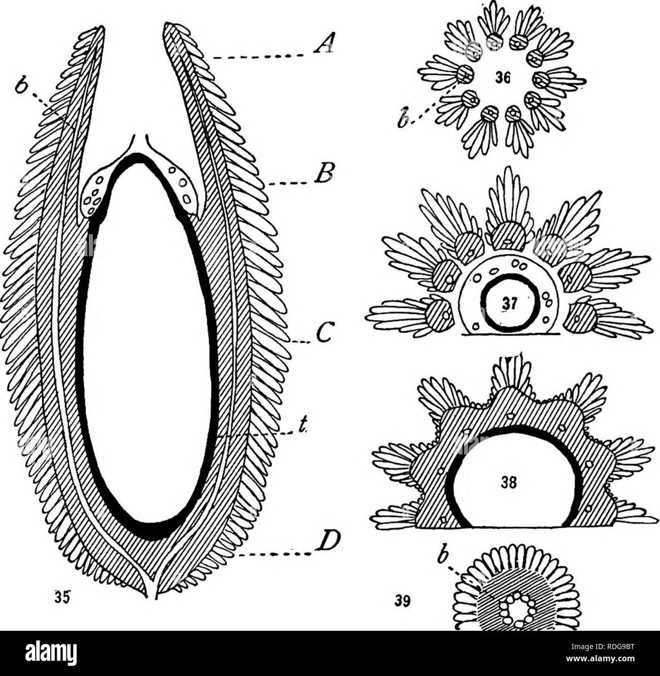
. Morphology of gymnosperms. Gymnosperms; Plant morphology. 36 MORPHOLOGY OF GYMNOSPERMS seem to be the units that make up the chambered integument ("canopy") of Lagenostoma, and this has suggested to Oliver the multiple origin of the integument. No stony layer is developed by the testa, and the distribution of the vascular strands is practically as in Lagenostoma, a single strand ascending deep in each rib and. Figs. 35-39.—Physostoma elegans: fig. 35, diagrammatic median longitudinal section; figs. 36-39, transverse sections at the levels A, B, C, D, respectively; b, vas- cular bundles; t, "tapetum."—After Oliver (85). passing on into the corresponding tentacle. The pollen chamber consists of the same circular crevice as in Lagenostoma, but the embryo sac develops into the nucellar projection (fig. 35), so that the pollen chamber really invests the upper end of the sac ("like the incurved bottom of a wine bottle"). The peripheral region of the nucellus, from the chalaza to the top of the embryo sac, is occupied by a remark- able "secretory zone, " made up of thin, oblong, tabular cells, and representing what Oliver regards as the retention of a feature present. Please note that these images are extracted from scanned page images that may have been digitally enhanced for readability - coloration and appearance of these illustrations may not perfectly resemble the original work.. Coulter, John Merle, 1851-1928; Chamberlain, Charles Joseph, b. 1863; Coulter, John Merle, 1851-1928. Morphology of spermatophytes. Part I. Gymnosperms. Chicago, University of Chicago Press