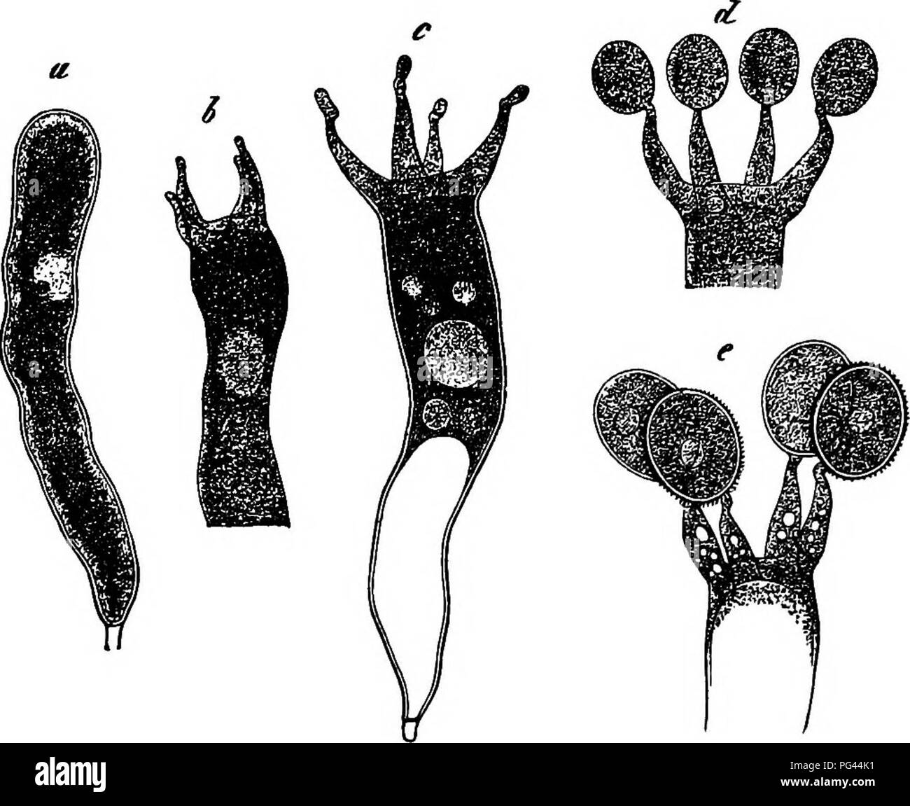
. Comparative morphology and biology of the fungi, mycetozoa and bacteria . Plant morphology; Fungi; Myxomycetes; Bacteriology. 64 DIVISION I.—GENERAL MORPHOLOGY. said; in Calocera and Dacryomyces they are cylindrical thin-walled cells rich in finely-granular protoplasm, which either entirely fills their inner space or is interrupted by vacuoles. It may be assumed that there is a nucleus always present, though in the smaller forms it has been looked for in vain up to the present time. Where it has been observed, as in Dacryomyces, Calocera, Corticium calceum, and especially in "the basidi
Image details
Contributor:
Central Historic Books / Alamy Stock PhotoImage ID:
PG44K1File size:
7.1 MB (314 KB Compressed download)Releases:
Model - no | Property - noDo I need a release?Dimensions:
1749 x 1428 px | 29.6 x 24.2 cm | 11.7 x 9.5 inches | 150dpiMore information:
This image is a public domain image, which means either that copyright has expired in the image or the copyright holder has waived their copyright. Alamy charges you a fee for access to the high resolution copy of the image.
This image could have imperfections as it’s either historical or reportage.
. Comparative morphology and biology of the fungi, mycetozoa and bacteria . Plant morphology; Fungi; Myxomycetes; Bacteriology. 64 DIVISION I.—GENERAL MORPHOLOGY. said; in Calocera and Dacryomyces they are cylindrical thin-walled cells rich in finely-granular protoplasm, which either entirely fills their inner space or is interrupted by vacuoles. It may be assumed that there is a nucleus always present, though in the smaller forms it has been looked for in vain up to the present time. Where it has been observed, as in Dacryomyces, Calocera, Corticium calceum, and especially in "the basidia of Corticium amorphum (Fig. 30) which become imm. in length, it is a spherical weakly refringent body (perhaps the nucleolus), lying in about the centre of the cell. It is not to be seen in the early states of the basidium, and it disappears when the formation of sterigmata commences. More exact investigation into its behaviour in spore-formation has yet to be made. When the basidium has reached its full size, the sterigmata make their appearance on its rounded apex as narrow subulate sprouts, and when they have arrived at a certain length their extremity, which up to this time is finely pointed, swells into a vesicle which gradually acquires the form, size, and structure of the mature spore. As the spore advances to maturity the protoplasm of the basidium passes into the swollen extremities, . Please note that these images are extracted from scanned page images that may have been digitally enhanced for readability - coloration and appearance of these illustrations may not perfectly resemble the original work.. Bary, A. de (Anton), 1831-1888; Garnsey, Henry E. F. (Henry Edward Fowler), 1826-1903; Balfour, Isaac Bayley, 1853-1922. Oxford : Clarendon Press