. Quarterly journal of experimental physiology and cognate medical sciences. lip of the fissure. In the contralateral cord-halfa faint scattered degeneration is still obvious occupying the usual pyra-midal-tract area. This latter degeneration is still detectible, althoughmuch less, at the 13th thoracic level, but lower than that is not recognis- The Excitable Cortex of the Cliimjxinzee, Oranjj-Utan, and (lorilla 185 able with certainty. In the lumbar enlarfjenient no defjeneration can bedetected. The ventral horn of ;;rey matter in the lower part ol the brachial en-lari^ement shows, especially

Image details
Contributor:
Reading Room 2020 / Alamy Stock PhotoImage ID:
2CEE4AXFile size:
7.2 MB (343.3 KB Compressed download)Releases:
Model - no | Property - noDo I need a release?Dimensions:
1561 x 1601 px | 26.4 x 27.1 cm | 10.4 x 10.7 inches | 150dpiMore information:
This image is a public domain image, which means either that copyright has expired in the image or the copyright holder has waived their copyright. Alamy charges you a fee for access to the high resolution copy of the image.
This image could have imperfections as it’s either historical or reportage.
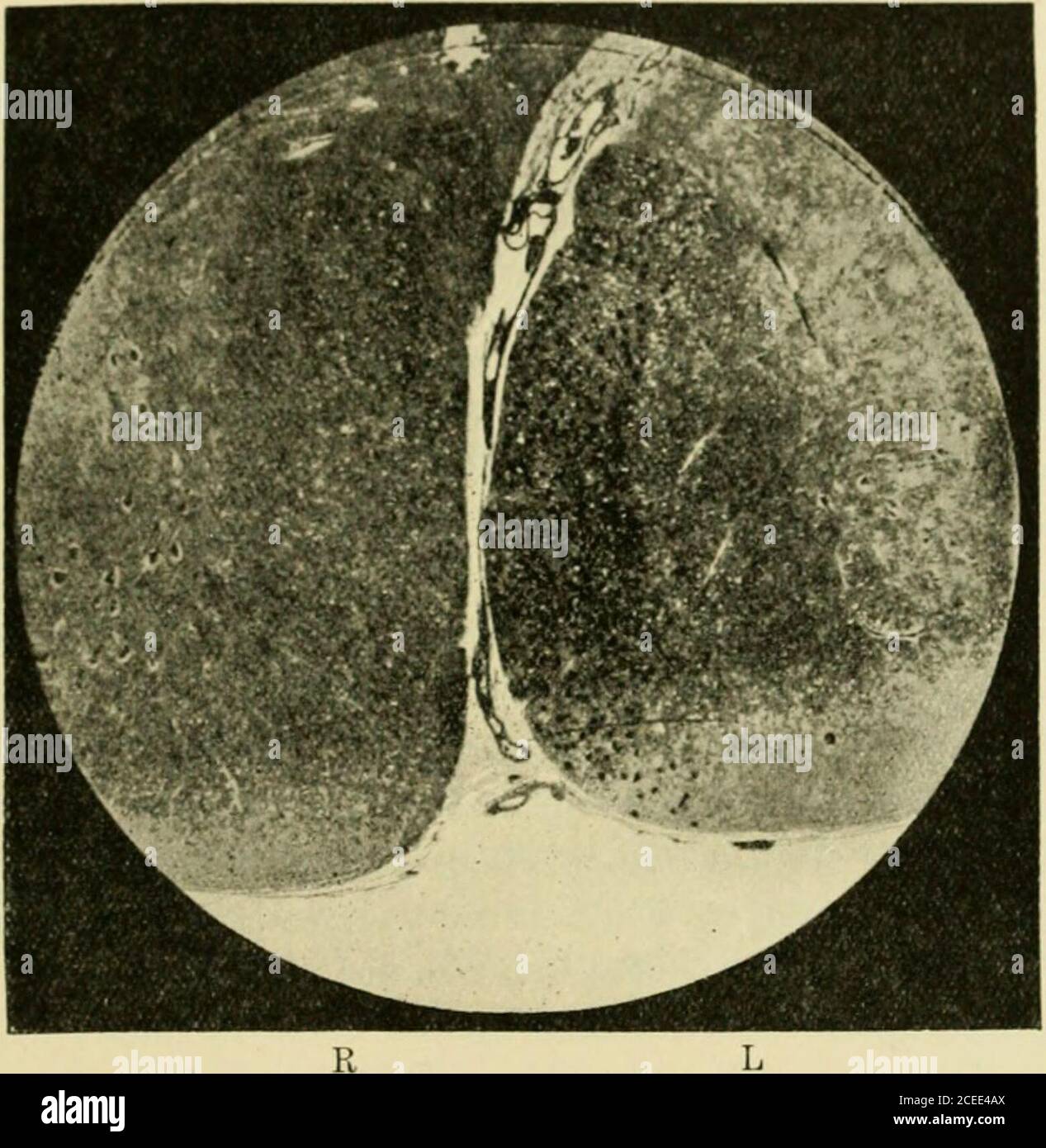
. Quarterly journal of experimental physiology and cognate medical sciences. lip of the fissure. In the contralateral cord-halfa faint scattered degeneration is still obvious occupying the usual pyra-midal-tract area. This latter degeneration is still detectible, althoughmuch less, at the 13th thoracic level, but lower than that is not recognis- The Excitable Cortex of the Cliimjxinzee, Oranjj-Utan, and (lorilla 185 able with certainty. In the lumbar enlarfjenient no defjeneration can bedetected. The ventral horn of ;;rey matter in the lower part ol the brachial en-lari^ement shows, especially in 7th and Sth cervical sei^ments, a markeddifierence between the rii, dit and left sides. ()n the right side, the wholeof the cross-area of ventral horn has scattered through it many degenerat-ing fibres of very minute size ; they give a peppered appearance to thegrey matter there, in contrast to the ordinary clean appearance of thecorresponding grey matter of the left half of the cord. The peppering isperhaps most marked in the dorsolateral and ventrolateral cell-group. Fig. 21.—Mici-ophotogram of direct pyramidal tract degenerated, along lip of ventral fissure, at 3rd cervical segment. Marchi preparation. Ablation-e.j)eriment 1. regions. It is certainly least in the medio-ventral cell-group. Sectionsstained with Marchi show these degenerated fibres in the grey matter butslightly, although, w^hen aware of them, one can detect the presence of anumber of them by that method. The degeneration in the ventral horn is, however, much better revealed by the Schafer (88) combination of theMarchi and Kulschitzky methods; the minute blue-black ring surroundingthe pale axis cylinder, w-hich many of the very small fibres in the greymatter give by that method, when seen in cross-section, is altered to aminute blob containing no axis cylinder. In other words, the fine col-laterals are degenerated, and their sheaths, with that element of it whichthe hsematoxylin stain after the mordant ting