BRAIN, MRI
RMID:Image ID:CRYJRX
Image details
Contributor:
BSIP SA / Alamy Stock PhotoImage ID:
CRYJRXFile size:
25 MB (1.4 MB Compressed download)Releases:
Model - no | Property - noDo I need a release?Dimensions:
2407 x 3630 px | 20.4 x 30.7 cm | 8 x 12.1 inches | 300dpiDate taken:
21 August 1995Photographer:
VEM / BSIPMore information:
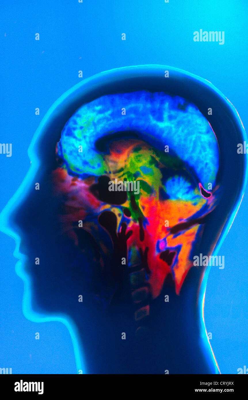
The two hemispheres of the brain are bridged by a thick band of nerve fibers known as the corpus callosum. The brain as a whole (encephalon) is composed of these areas along with the brain stem (the prolongation of the spinal cord), and the cerebellum (a sort of branch circuit located in the posterior region of the skull). MRI imagery, which does not produce any radiation, is particularly useful for brain exams, especially those targeting the posterior cranial fossa and the white matter. Seen here, a normal, healthy brain.