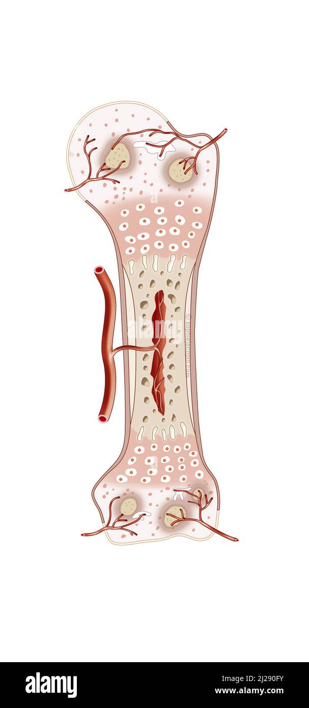
Newborn bone tissue
RMID:Image ID:2J290FY
Image details
Contributor:
BSIP SA / Alamy Stock PhotoImage ID:
2J290FYFile size:
59.9 MB (526.1 KB Compressed download)Releases:
Model - no | Property - noDo I need a release?Dimensions:
3130 x 6694 px | 26.5 x 56.7 cm | 10.4 x 22.3 inches | 300dpiDate taken:
17 April 2021Location:
FrancePhotographer:
JACOPIN / BSIPMore information:
Newborn bone tissue, bone development, cartilage structure, pediatrics. This illustration shows the anatomy of a humerus in frontal section. From birth to childhood, the bone structure is cartilaginous and flexible. The center of the bone presents a vascularized medullary cavity. Compact bone is formed around this cavity (beige area in the center). At each end of this area is the epiphyseal cartilage (in pink). This area is more important in infants than in children. It has disappeared in the adult. Then the ends of the bone (epiphyses) are very vascularized.