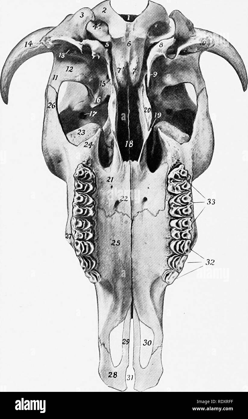
. The anatomy of the domestic animals . Veterinary anatomy. 136 SKELETON OF THE OX length of the skull, and all of the roof of the cranium. The posterior borders form with the parietals a large central frontal eminence (Torus frontalis), the highest point of the skull. At the junction of the posterior and the lateral border is the processus comus or "horn core," for the support of the horn. These proc-. FiG. 134.—Skull OF Ox, without Mandible; Ventral View. 1, Foramen magnum; 2, occipital condyle; 3, paramastoid process; 4, condyloid foramen; 5, foramen lacerum; 6, basilar part of oc
Image details
Contributor:
The Book Worm / Alamy Stock PhotoImage ID:
RDXRFFFile size:
7.2 MB (307 KB Compressed download)Releases:
Model - no | Property - noDo I need a release?Dimensions:
1258 x 1987 px | 21.3 x 33.6 cm | 8.4 x 13.2 inches | 150dpiMore information:
This image is a public domain image, which means either that copyright has expired in the image or the copyright holder has waived their copyright. Alamy charges you a fee for access to the high resolution copy of the image.
This image could have imperfections as it’s either historical or reportage.
. The anatomy of the domestic animals . Veterinary anatomy. 136 SKELETON OF THE OX length of the skull, and all of the roof of the cranium. The posterior borders form with the parietals a large central frontal eminence (Torus frontalis), the highest point of the skull. At the junction of the posterior and the lateral border is the processus comus or "horn core, " for the support of the horn. These proc-. FiG. 134.—Skull OF Ox, without Mandible; Ventral View. 1, Foramen magnum; 2, occipital condyle; 3, paramastoid process; 4, condyloid foramen; 5, foramen lacerum; 6, basilar part of occipital bone; 7, 1', basilar tubercles; 8, bulla ossea; 9, foramen ovale (concealed by muscular proc- ess) ; 10, meatus acusticus externus; 11, z^'gomatic process of temporal bone, 12, condyle of same; 13, external opening of temporal canal; 14, processus cornus; 15, muscular process of temporal bone; 16, pterygoid crest; 17, orbital open- ing of supraorbital canal; 18, choante or posterior nares; 19, hamulus of pterygoid bone; 20, crest formed by pterygoid processes of sphenoid and palatine bones; 21, horizontal part of palatine bone; 22, anterior palatine foramen; 23, lacrimal bulla; 24, maxillary tuberosity; 25, palatine process of maxilla; 26, zygomatic process of malar bone; 27, facial tuberosity: 28, body of premaxilla; 29, palatine process of same; 30, palatine fissure; 31, incisive fissure; 32, premolars; 33, molars. esses are of elongated conical form, and vary greatly in size, length, curvature, and direction. The external surface is rough and porous, marked by numerous grooves and foramina; in the fresh state it is covered by the corium of the horn. The base has a constriction, the neck. The interior is excavated to form a number of irregular spaces, partially divided by bony septa, and communicating with the. Please note that these images are extracted from scanned page images that may have been digitally enhanced for readability - coloration and appearance of the