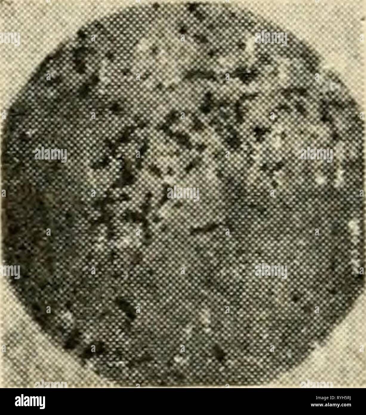
The electron microscope, its development, present performance and future possibilities electronmicrosco00gabo Year: 1948 ^ (a) B. Subtilis • (b) Typhoid Fig. 28. Typhoid germ and Bacillus Subtilis electron and light micrographs density. This for the time being is still a difficult and rather uncertain process, and in the case of small objects their theory requires correction for the reasons described in chapter 6. The biologist will obtain in most cases sufficient information from a few micrographs which show the bacilli in different positions or directly from stereoscopic electron micro
Image details
Contributor:
Bookend / Alamy Stock PhotoImage ID:
RYH5RJFile size:
5.7 MB (272.6 KB Compressed download)Releases:
Model - no | Property - noDo I need a release?Dimensions:
1374 x 1455 px | 23.3 x 24.6 cm | 9.2 x 9.7 inches | 150dpiMore information:
This image is a public domain image, which means either that copyright has expired in the image or the copyright holder has waived their copyright. Alamy charges you a fee for access to the high resolution copy of the image.
This image could have imperfections as it’s either historical or reportage.
The electron microscope, its development, present performance and future possibilities electronmicrosco00gabo Year: 1948 ^ (a) B. Subtilis • (b) Typhoid Fig. 28. Typhoid germ and Bacillus Subtilis electron and light micrographs density. This for the time being is still a difficult and rather uncertain process, and in the case of small objects their theory requires correction for the reasons described in chapter 6. The biologist will obtain in most cases sufficient information from a few micrographs which show the bacilli in different positions or directly from stereoscopic electron micrographs. By these means it is possible to overcome the disadvantage of electron micrographs which like X-ray diagrams have very great depth of focus and make it difficult to locate details in depth. This The bacteriophages, or bacterial viruses, of which three strains have been identified under the electron microscope, are the smallest known liv- ing organisms. For some time it was doubtful whether they could be classed as such. The strain here shown, Bacteriophage anti-coli PC, is the largest of the three. The 'head' has a diameter of about 800 A (0.08 microns), the 'tail' has a length of about 1, 300 A. One particle of phage is sufficient to originate the lysis of a bacterial cell, and during the lysis an average of about a hundred new phage particles are generated. This photograph shows the phage particles swimming toward the bacteria. They were immobilized by drying five minutes after a suspension of the bacteria was injected with a drop of diluted phage solution. (By courtesy of Dr. V. K. Zworykin, R.C.A.)