An atlas of human anatomy for students and physicians . S.vc. Topographical Anatomy of the Thoracic Organs and of the Viscera in the Upper Part of the Abdominal Cavity. 480 TOPOGRAPHICAL ANATOMY OF THE THORACIC AND ABDOMINAL VISCERA Diaphragm (right inner crus or pillar)Diaphragma (crus mediale dextrum)Right kidney (upper extremity)Ken dexter (extremitas superior)Retroperitoneal space —Spatium retroperitoneal Peritoneal cavity—Cavum peritonaei Parietal peritonseum —Peritoneum pariet. Diaphragm (costal portion) Diaphragma (pars costalis)Pleural cavity (phrenocostal ordiaphragmaticocostal supple
Image details
Contributor:
The Reading Room / Alamy Stock PhotoImage ID:
2AJB95NFile size:
7.1 MB (554.4 KB Compressed download)Releases:
Model - no | Property - noDo I need a release?Dimensions:
1552 x 1610 px | 26.3 x 27.3 cm | 10.3 x 10.7 inches | 150dpiMore information:
This image is a public domain image, which means either that copyright has expired in the image or the copyright holder has waived their copyright. Alamy charges you a fee for access to the high resolution copy of the image.
This image could have imperfections as it’s either historical or reportage.
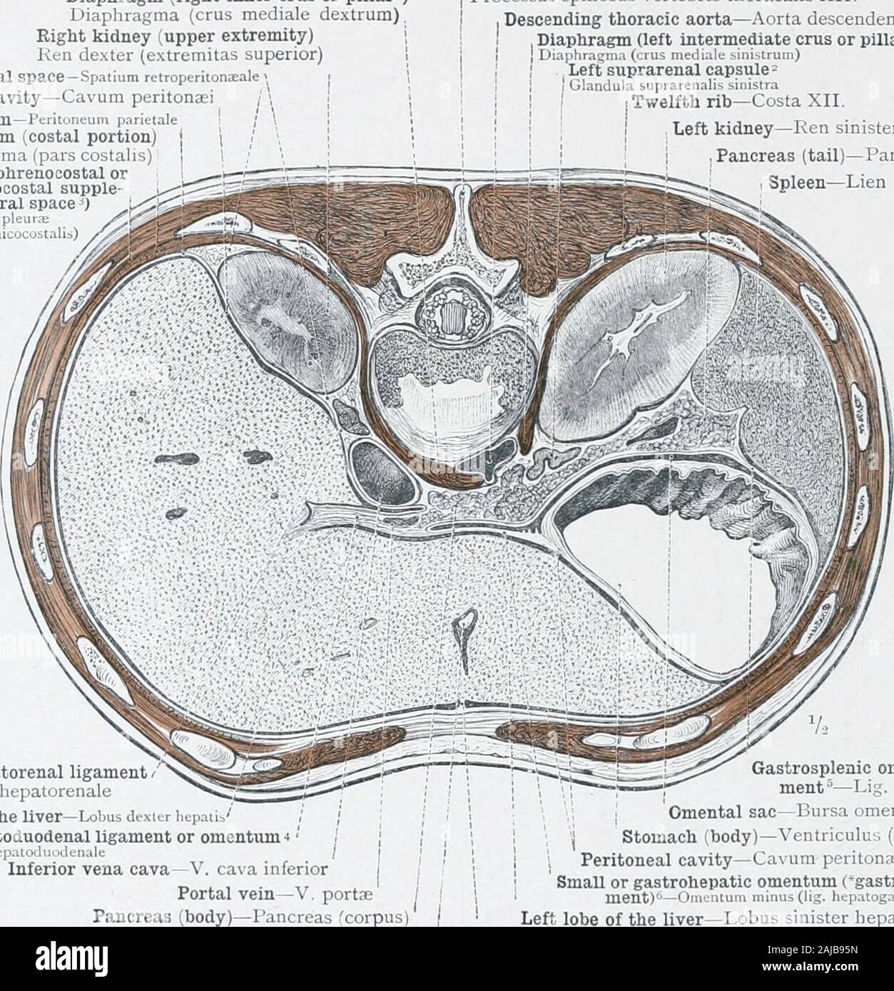
An atlas of human anatomy for students and physicians . S.vc. Topographical Anatomy of the Thoracic Organs and of the Viscera in the Upper Part of the Abdominal Cavity. 480 TOPOGRAPHICAL ANATOMY OF THE THORACIC AND ABDOMINAL VISCERA Diaphragm (right inner crus or pillar)Diaphragma (crus mediale dextrum)Right kidney (upper extremity)Ken dexter (extremitas superior)Retroperitoneal space —Spatium retroperitoneal Peritoneal cavity—Cavum peritonaei Parietal peritonseum —Peritoneum pariet. Diaphragm (costal portion) Diaphragma (pars costalis)Pleural cavity (phrenocostal ordiaphragmaticocostal supplemental pleural spaced) Cavum pie u u(sinus phrsnicoLo t , !?=) Spinous process of the twelfth dorsal vertebra Processus spinosus vertebrae thoracalis XII. Descending thoracic aorta—Aorta descendens, Diaphragm (left intermediate crus or pillar) I DiaphraKraa (crus mediale sinistrum) Left suprarenal capsule= Glandula suprarenalis sinistra [Twelfth rib—Costa XII. I , Left kidney—Ren sinister , Pancreas (tail)—Pancreas (cauda)Spleen—Lien. Hepatorenal ligament Lig. hepaiorenaieRight lobe of the liver—Lobus dexier liepatis ; ; ?Hepatoduodenal ligament or omentum 4 , ?Lig. liepatoJu.denale / Inferior vena cava—V. caa inferior Portal vein—V. portsePancreas (body)—Pancreas (corpus Round ligament of the liverLig. teres hepatis See Appendix, note 37. 2 Called also supraraialiody, or adrenal. 5 The gastros^icnic omentum is connected below with the great omentum, 6 See Appendix, note 42. Gastiosplenic omentum or liga-ment —L, g. gastrolienale, ; > Omental sac—Bursa omentalis j Stomach (body)—Ventriculus (corpus) Peritoneal cavity—Cavum peritona;! SmaU or gastrohepatic omentum Cgastrohepatic liga-ment)-OuKiuum minus (lij. hepatogastricuni) Left lobe of the liver—Lobus sinister hepatis Falciform, broad, or suspensory ligament of the liver Lig. falciforme hepatis See Appendix, note 36. 4 See Appendix, note 4=. often regarded as a part of it.—Tr. Fig, 8io.-