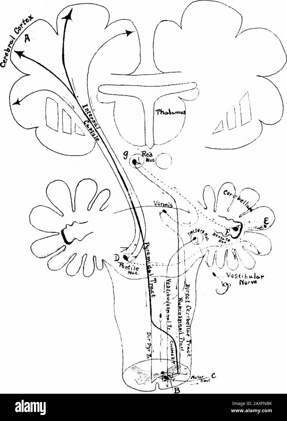
A manual of anatomy . through the corona radiata intothe internal capsule occupying the middle portion thereof; they enterthe crusta of the crus cerebri, then the tegmentum of the pons andthe ventral area of the oblongata; in these three regions some of itsfibers pass to the cerebral nerve nuclei of origin. At the caudal endof the oblongata 85 to 90 per cent, of the fibers decussate to the op-posite side of the spinal cord as the crossed pyramidal tract and thenend at various levels around the cells of the ventral horn. Theremaining fibers continue down the same side of the spinal cord, asthe

Image details
Contributor:
The Reading Room / Alamy Stock PhotoImage ID:
2AXFNBKFile size:
7.2 MB (226.7 KB Compressed download)Releases:
Model - no | Property - noDo I need a release?Dimensions:
1351 x 1850 px | 22.9 x 31.3 cm | 9 x 12.3 inches | 150dpiMore information:
This image is a public domain image, which means either that copyright has expired in the image or the copyright holder has waived their copyright. Alamy charges you a fee for access to the high resolution copy of the image.
This image could have imperfections as it’s either historical or reportage.
A manual of anatomy . through the corona radiata intothe internal capsule occupying the middle portion thereof; they enterthe crusta of the crus cerebri, then the tegmentum of the pons andthe ventral area of the oblongata; in these three regions some of itsfibers pass to the cerebral nerve nuclei of origin. At the caudal endof the oblongata 85 to 90 per cent, of the fibers decussate to the op-posite side of the spinal cord as the crossed pyramidal tract and thenend at various levels around the cells of the ventral horn. Theremaining fibers continue down the same side of the spinal cord, asthe direct pyramidal tract, to various levels in the cervical and upper THE MOTOR PATHWAY 417 thoracic region and then pass through the ventral, or white com-missure, to end in the ventral horn of the opposite side. Ultimatelyall fibers decussate before they end. This ends the first neuron. Thesecond neuron comprises the cells of the ventral horn and theirprocesses that form the motor root of the spinal nerves, on the one. F, G. 302. Diagram of the neurons in the direct and indirect motor pathways and the con-nections of the cerebellum with the brain stem and the spinal cord. Direct.—Neuron I. .4toB; neuron 2, B to C. Indirect.—Neuron I, .-1 fcerebral cortex) to D; 2, D to E; 3, £ to F; 4.Fto g; 5, s to B; 6, B to C. hand, and in the case of the cerebral nerves comprises the cells ofthe various nuclei of origin and their processes that form the motorportion of the cerebral nerves. These axones pass out of the graysubstance, become myehnated and ultimately end directly in a vol-untary striated muscle fiber. This is the end of the second neuron.First new on, a pj^ramidal cell in the motor cortex of the cerebrum 4i8 THE NERVE SYSTEM and its axone that forms a part of the pyramidal tract and thatends in a cerebral nerve nucleus, or the ventral gray of the spinalcord. Second neuron, the cell in the nucleus of origin or in the ventral