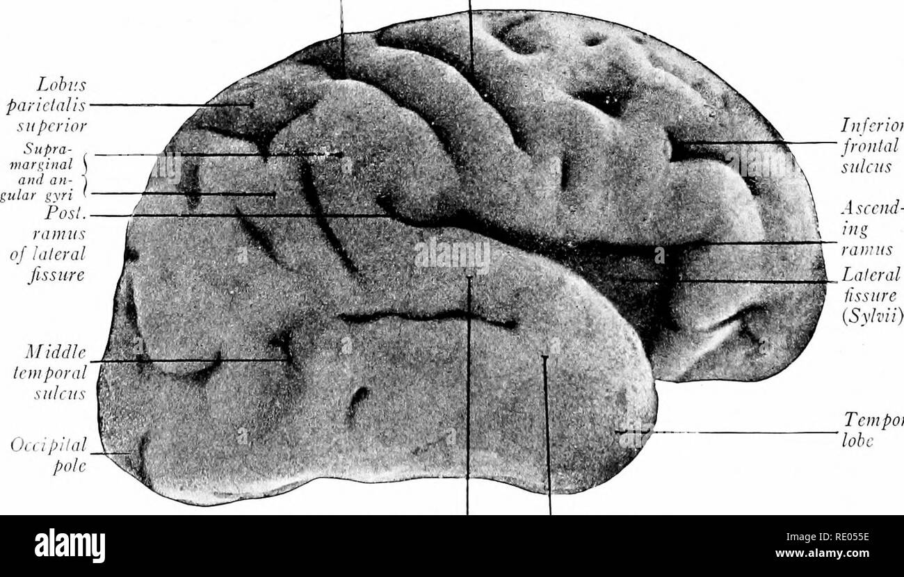
. A laboratory manual and text-book of embryology. Embryology. Insula Lai. olfactory gyrus Gyrus ambiens Gyrus semilunaris Oliva Fig. 336.—Ventral view of the brain of a 100 mm. embryo showing development of the rhinencephalon (Kollmann). Sulcus postcentral is Sulcus centralis Middle temporal sulcus Occipital pole Inferior frontal sulcus Ascend-. Temporal lobe Superior temporal gyrus Middle lemporal gyrus Fig. 337. —Lateral view of the right cerebral hemisphere, from a seven months' fetus (Kollmann).. Please note that these images are extracted from scanned page images that may have been digit
Image details
Contributor:
The Book Worm / Alamy Stock PhotoImage ID:
RE055EFile size:
7.1 MB (424.1 KB Compressed download)Releases:
Model - no | Property - noDo I need a release?Dimensions:
2094 x 1193 px | 35.5 x 20.2 cm | 14 x 8 inches | 150dpiMore information:
This image is a public domain image, which means either that copyright has expired in the image or the copyright holder has waived their copyright. Alamy charges you a fee for access to the high resolution copy of the image.
This image could have imperfections as it’s either historical or reportage.
. A laboratory manual and text-book of embryology. Embryology. Insula Lai. olfactory gyrus Gyrus ambiens Gyrus semilunaris Oliva Fig. 336.—Ventral view of the brain of a 100 mm. embryo showing development of the rhinencephalon (Kollmann). Sulcus postcentral is Sulcus centralis Middle temporal sulcus Occipital pole Inferior frontal sulcus Ascend-. Temporal lobe Superior temporal gyrus Middle lemporal gyrus Fig. 337. —Lateral view of the right cerebral hemisphere, from a seven months' fetus (Kollmann).. Please note that these images are extracted from scanned page images that may have been digitally enhanced for readability - coloration and appearance of these illustrations may not perfectly resemble the original work.. Prentiss, Charles William, 1874-1915. Philadelphia, London, W. B. Saunders