. Comparative anatomy of vertebrates. Anatomy, Comparative; Vertebrates. INTRODUCTION 11 allantois. The latter becomes attached to a definite region of the uterine wall, and from it vascular processes or villi arise, so that the foetal and maternal blood-vessels come into very close relations with one another. Thus an allantoic placenta is Pa PC(CLf). Dv FIG. 9.—DIAGRAMMATIC .SECTION THROUGH THE HUMAN GRAVID UTERUS. A, aorta ; A, A, A, the cavity of the amnion filled with fluid : in the interior of the amnion is seen the embryo suspended by the twisted umbilical cord; Al, allantoic (umbilical)
Image details
Contributor:
The Book Worm / Alamy Stock PhotoImage ID:
REFE12File size:
7.1 MB (325.4 KB Compressed download)Releases:
Model - no | Property - noDo I need a release?Dimensions:
1495 x 1671 px | 25.3 x 28.3 cm | 10 x 11.1 inches | 150dpiMore information:
This image is a public domain image, which means either that copyright has expired in the image or the copyright holder has waived their copyright. Alamy charges you a fee for access to the high resolution copy of the image.
This image could have imperfections as it’s either historical or reportage.
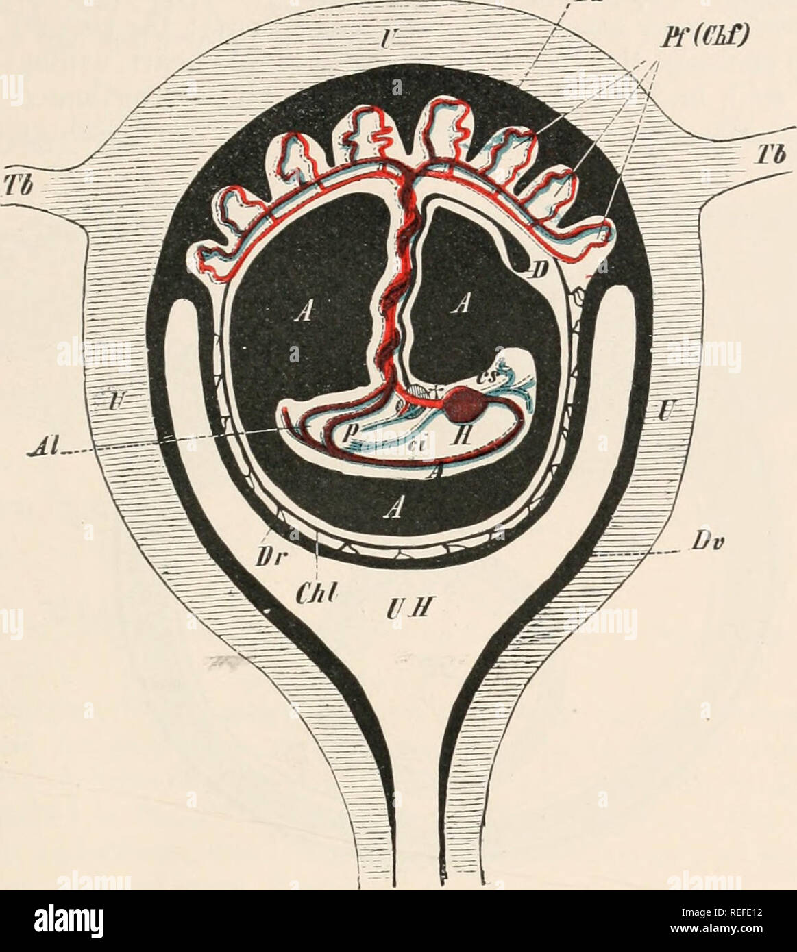
. Comparative anatomy of vertebrates. Anatomy, Comparative; Vertebrates. INTRODUCTION 11 allantois. The latter becomes attached to a definite region of the uterine wall, and from it vascular processes or villi arise, so that the foetal and maternal blood-vessels come into very close relations with one another. Thus an allantoic placenta is Pa PC(CLf). Dv FIG. 9.—DIAGRAMMATIC .SECTION THROUGH THE HUMAN GRAVID UTERUS. A, aorta ; A, A, A, the cavity of the amnion filled with fluid : in the interior of the amnion is seen the embryo suspended by the twisted umbilical cord; Al, allantoic (umbilical) arteries ; Chi, chorion Izeve ; D, the remains of the yolk-sack (umbilical vesicle) ; Dr, decidua reflexa; Dv, decidua vera, which at Pu passes into the uterine portion of the placenta ; H, heart; Pf, f<etal portion of the placenta (chorion frondosum, Chf) : Tb, Fallopian tube; U, uterine wall; UH, uterine cavity ; <:i, postcaval, cs, precaval, and p. portal vein ; f, the liver, perforated by the umbilical vein. formed, which serves both for the respiration and nutrition of the foetus (Fig. 9). The following important points must now be noted as regards the structure of the Vertebrate body. After the above-mentioned main organs have appeared, a smaller dorsal neural tube and a larger ventral visceral tube are seen to extend longitudinally through the body, and between the two is a rod-like support- ing structure, the notochord (p. 6), which forms the primary. Please note that these images are extracted from scanned page images that may have been digitally enhanced for readability - coloration and appearance of these illustrations may not perfectly resemble the original work.. Wiedersheim, Robert, 1848-1923; Parker, W. N. (William Newton), d. 1923. London, Macmillan and co. , limited