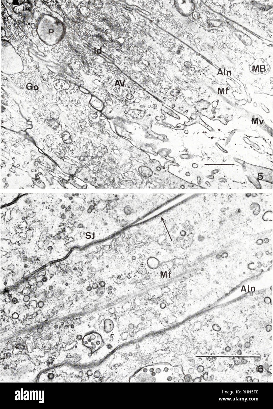
. The Biological bulletin. Biology; Zoology; Biology; Marine Biology. 214 HERBERT E. POTSWALD T7- *3&**^»''^f' • -. FIGURE 5. Apical region of the pore epithelium of a brooding operculum showing that each cell has an apical Golgi complex (Go), multivesicular bodies (MB), pigment granules (P), lateral interdigitations (Id), apical vesicles (AV), microvilli (Mv), and longi- tudinal bundles of microfilaments (Mf). Note also that the epithelial cells are separated by apical intercellular spaces (AIn). Scale equals 1 /*. FIGURE 6. Similar to Figure 5 but at higher magnification, showing septate
Image details
Contributor:
Library Book Collection / Alamy Stock PhotoImage ID:
RHN5TEFile size:
7.1 MB (610.9 KB Compressed download)Releases:
Model - no | Property - noDo I need a release?Dimensions:
1337 x 1868 px | 22.6 x 31.6 cm | 8.9 x 12.5 inches | 150dpiMore information:
This image is a public domain image, which means either that copyright has expired in the image or the copyright holder has waived their copyright. Alamy charges you a fee for access to the high resolution copy of the image.
This image could have imperfections as it’s either historical or reportage.
. The Biological bulletin. Biology; Zoology; Biology; Marine Biology. 214 HERBERT E. POTSWALD T7- *3&**^»''^f' • -. FIGURE 5. Apical region of the pore epithelium of a brooding operculum showing that each cell has an apical Golgi complex (Go), multivesicular bodies (MB), pigment granules (P), lateral interdigitations (Id), apical vesicles (AV), microvilli (Mv), and longi- tudinal bundles of microfilaments (Mf). Note also that the epithelial cells are separated by apical intercellular spaces (AIn). Scale equals 1 /*. FIGURE 6. Similar to Figure 5 but at higher magnification, showing septate junctions (SJ), apical intercellular spaces (AIn), longitudinal bundles of microfilaments (Mf), and circumferential network of microfilaments adjacent to the apical plastnalemma (arrow). Scale equals 1 /*.. Please note that these images are extracted from scanned page images that may have been digitally enhanced for readability - coloration and appearance of these illustrations may not perfectly resemble the original work.. Marine Biological Laboratory (Woods Hole, Mass. ); Marine Biological Laboratory (Woods Hole, Mass. ). Annual report 1907/08-1952; Lillie, Frank Rattray, 1870-1947; Moore, Carl Richard, 1892-; Redfield, Alfred Clarence, 1890-1983. Woods Hole, Mass. : Marine Biological Laboratory