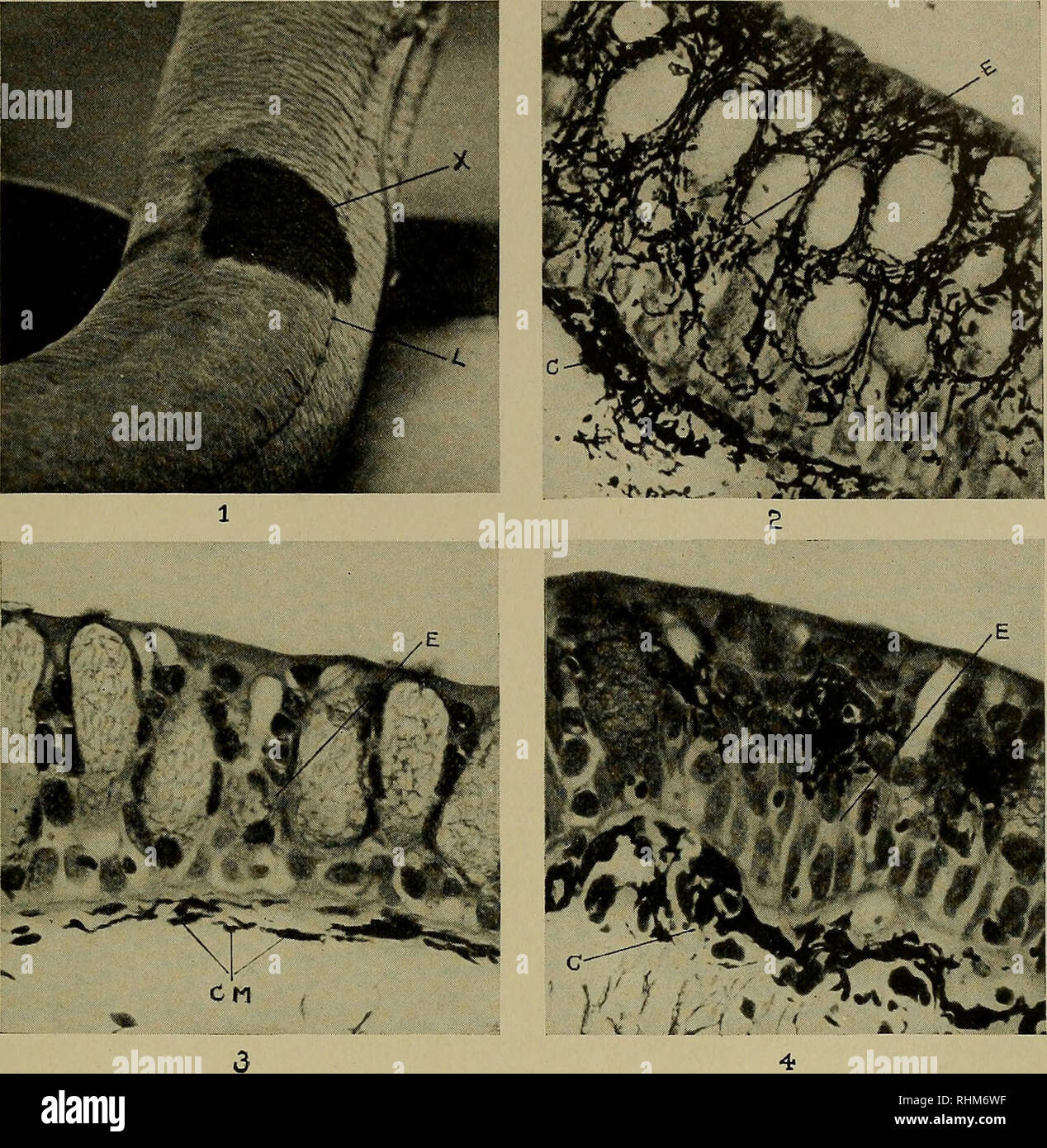
. The Biological bulletin. Biology; Zoology; Marine biology. CUTANEOUS MELANOSIS IN LUNGFISHES 283. Plate I EXPLANATION OF PLATE I Fig. 1. Lepidosiren paradoxa (lungfish A) with pigmented patch {X) in the right dorsal region encroaching on the lateral line (L). Fig. 2. Photomicrograph of melanotic tissue removed from lungfish A, showing a hyperplasia of melanophores infiltrating thickened epidermis (£). Numerous melanophores in the corium (C). Rogers' silver stain, counterstained faintly with erythrosin. X 250. Fig. 3. Normal skin removed from lungfish B. E. epidermis; CM. corial melanophores.
Image details
Contributor:
Library Book Collection / Alamy Stock PhotoImage ID:
RHM6WFFile size:
7.1 MB (629.5 KB Compressed download)Releases:
Model - no | Property - noDo I need a release?Dimensions:
1561 x 1600 px | 26.4 x 27.1 cm | 10.4 x 10.7 inches | 150dpiMore information:
This image is a public domain image, which means either that copyright has expired in the image or the copyright holder has waived their copyright. Alamy charges you a fee for access to the high resolution copy of the image.
This image could have imperfections as it’s either historical or reportage.
. The Biological bulletin. Biology; Zoology; Marine biology. CUTANEOUS MELANOSIS IN LUNGFISHES 283. Plate I EXPLANATION OF PLATE I Fig. 1. Lepidosiren paradoxa (lungfish A) with pigmented patch {X) in the right dorsal region encroaching on the lateral line (L). Fig. 2. Photomicrograph of melanotic tissue removed from lungfish A, showing a hyperplasia of melanophores infiltrating thickened epidermis (£). Numerous melanophores in the corium (C). Rogers' silver stain, counterstained faintly with erythrosin. X 250. Fig. 3. Normal skin removed from lungfish B. E. epidermis; CM. corial melanophores. Hematoxylin and eosin. X 250. Fig. 4. Tissue removed from melanotic patch of lungfish B. Mild increase of melanophores in corium C and a moderate infiltration of epidermis E by melanophores. Hematoxylin and eosin. X 250.. Please note that these images are extracted from scanned page images that may have been digitally enhanced for readability - coloration and appearance of these illustrations may not perfectly resemble the original work.. Lillie, Frank Rattray, 1870-1947; Moore, Carl Richard, 1892-; Redfield, Alfred Clarence, 1890-; Marine Biological Laboratory (Woods Hole, Mass. ); Marine Biological Laboratory (Woods Hole, Mass. ). Annual report; HighWire Press. Lancaster, Pa. [etc. ] : Lancaster Press, inc. [etc. ]